
Ed Boyden
@eboyden3
Followers
27K
Following
6K
Media
303
Statuses
4K
Y. Eva Tan Professor in Neurotechnology, MIT. Investigator, HHMI. Leader, Synthetic Neurobiology Group. Inventor. Entrepreneur.
MIT and HHMI, Cambridge, MA
Joined November 2008

I’m so excited to share my first-author PhD paper, out today in @NeuroCellPress as a NeuroResource! A huge thanks to all of my co-authors, and of course to my wonderful mentors Guoping Feng, @fennamk, and @eboyden3.
cell.com
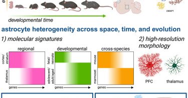
In this NeuroResource, Schroeder et al. present a transcriptomic atlas across brain regions and developmental time points in mouse and marmoset. Detailed analysis focused on astrocytes revealed that...
6
9
92
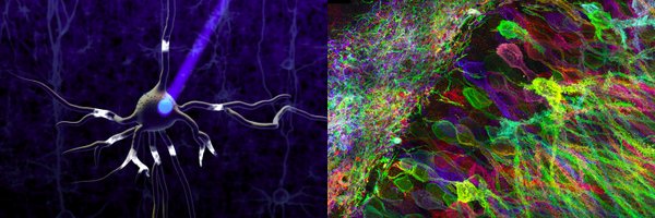
Diatoms – microscopic algae found almost everywhere there’s water – make up ~25% of Earth’s annual oxygen production. EMBL scientists have found a way to reveal their inner structures by combining cryo-fixation with ultrastructural expansion microscopy. https://t.co/BxmvHag8Kb
5
12
26

Applying new tools to entire brains, starting with C. elegans, offers the opportunity to uncover how molecules work together to generate neural physiology, and how neurons generate behavior, write @eboyden3 and @koerding. https://t.co/TMjnGwbAPh

thetransmitter.org
Applying new tools to entire brains, starting with C. elegans, offers the opportunity to uncover how molecules work together to generate neural physiology and how neurons work together to generate…
3
23
77

Thank you to everyone who joined the Neurotechnology Workshop – BCI, WBE & AGI with us, this weekend! @eboyden3, @patrickmineault, @Andrew_C_Payne, @mardinly, @michaelandregg, @stardazed0, @EscolaSean70058, @JacquesCarolan, @halehf, @AToliasLab, @BobbyKasthuri, @gkreiman,
4
15
80

Keynote talk @NSTumorSection @CNS_Update by @eboyden3 - a tour de force from #expansionmicroscopy to #optogenics and beyond - what a treat to learn about the latest in #neurotechnologies - thank you @eboyden3 for sharing with this group of #braintumor #neurosurgeons
2
4
11
Paper: https://t.co/vaYDVtnsAX We partnered with @SGRodriques (Francis Crick Institute, now Future House), @eboyden3 (MIT and HHMI), and @jmrko (MPI) for this study.

biorxiv.org
Mapping nanoscale neuronal morphology with molecular annotations is critical for understanding healthy and dysfunctional brain circuits. Current methods are constrained by image segmentation errors...
2
7
36
@E11BIO is excited to unveil PRISM technology for mapping brain wiring with simple light microscopes. Today, brain mapping in humans and other mammals is bottlenecked by accurate neuron tracing. PRISM uses molecular ID codes and AI to help neurons trace themselves. We discovered
36
111
369

Today, with @E11BIO, I am excited to announce PRISM, a new, scalable technology for mapping brain circuits. Within 10 years, technologies like PRISM will enable us to understand fundamental brain functions, fight brain disorders, and maybe even achieve upload. Enormous

@E11BIO is excited to unveil PRISM technology for mapping brain wiring with simple light microscopes. Today, brain mapping in humans and other mammals is bottlenecked by accurate neuron tracing. PRISM uses molecular ID codes and AI to help neurons trace themselves. We discovered
1
22
137

We are thrilled to announce that Sven Dorkenwald @sdorkenw will join the McGovern Institute faculty in January 2026! 🎉 Sven uses advanced computational methods to build and analyze comprehensive maps of the brain. @mitbrainandcog @ScienceMIT
https://t.co/8FDAr17WWP

mcgovern.mit.edu
The McGovern Institute and the Department of Brain and Cognitive Sciences are pleased to announce the appointment of Sven Dorkenwald as an assistant professor starting in January 2026. A trailblazer...
1
2
29
As part of our recent 25th anniversary celebration, our Director Bob Desimone reflected on some of our greatest accomplishments and described what we might expect to see from the McGovern Institute in the next decade. Watch his talk below 👇#TalkTuesday
https://t.co/PfydLfG7My
0
4
14

"Academic research groups and startups are essential drivers of scientific progress. But some projects, like the Hubble Space Telescope or the Human Genome Project, are too big for any one academic lab or loose consortium. They’re also not immediately profitable enough for

news.mit.edu
Developed by former MIT researchers, focused research organizations (FROs) undertake large research efforts and have begun to contribute to scientific advances.
1
7
42
MIT News featured the origin story of Focused Research Organizations and @E11BIO coming out of @eboyden3's lab with @AdamMarblestone @SGRodriques @Andrew_C_Payne @tkalil2050
https://t.co/KWPmtsVufp
news.mit.edu
Developed by former MIT researchers, focused research organizations (FROs) undertake large research efforts and have begun to contribute to scientific advances.
0
6
26

It feels that biographies of 19th century and early 20th century figures will in some regard never be matched: because the culture was one of abundant letter-writing, we often get unparalleled insight into the subjects' frames of mind and thought processes. Today, of course, we
132
99
2K

Researchers combined in situ sequencing with expansion microscopy to get more spatial insight into how stretches of the genome interact with proteins in the jam-packed nucleus.
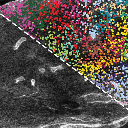
cen.acs.org
Researchers combine expansion microscopy with sequencing to probe chromatin organization
0
1
6

Super excited that Expansion In Situ Genome Sequencing (ExIGS) is out now in @ScienceMagazine ! Check out the paper to see what can be revealed by sequencing the genome in its native position and high-res imaging its interactions with nearby proteins. https://t.co/weVnIz7RD1
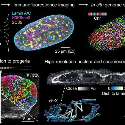
science.org
Microscopy and genomics are used to characterize cell function, but approaches to connect the two types of information are lacking, particularly at subnuclear resolution. Here, we describe expansion...
1
14
56

A new combination technique enables DNA sequencing and high-resolution imaging in intact cells — offering new insights into progeria and aging. https://t.co/8k1cD56eJ1
@JD_Buenrostro @insitubiology @ajaylabade31 @CarolineComenho @z_chiang @ScienceMagazine
4
71
245

The peer-reviewed version of expansion in situ genome sequencing is now out in Science! The news is bittersweet – when we first revealed this last September, I never guessed it would be my final paper in academia, but a lot has changed. A few parting thoughts:
18
298
1K

ExIGS advances the field of "expansion genomics," dramatically reshaping how we see and understand genome organization by bridging scales, uncovering hidden details, and asking entirely new biological questions.#ExpansionGenomics #SpatialGenomics
1
1
9
Thrilled to announce that our work on Expansion In Situ Genome Sequencing (ExIGS) is now published in @ScienceMagazine! ExIGS allows us to sequence DNA and image proteins with super-resolution directly within single cells. https://t.co/CEIsZdFrPU
science.org
Microscopy and genomics are used to characterize cell function, but approaches to connect the two types of information are lacking, particularly at subnuclear resolution. Here, we describe expansion...
9
46
201