carolinebsirini, MD
@cbsirini
Followers
902
Following
974
Media
38
Statuses
405
Cytopathology fellow @Emorypathology/ former GI path fellow and resident at @pathology_URMC
Joined November 2017
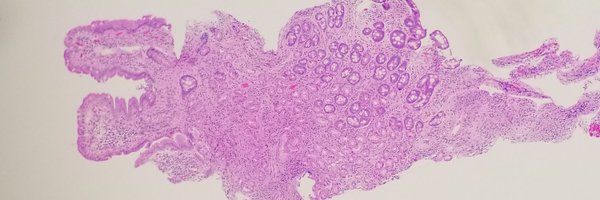
My beautiful CNSET case!

15 yo f presented to ED w/acute onset of RLQ abd pain. CT scan revealed an incidental 14 cm heterogenous mass in the right lobe of the liver. Case by Caroline Bsirini MD, Karen Vanderbilt BS, Lee Ann Hellman PA (ASCP), Michael G. Drage MD, PhD, #Liverpath
https://t.co/NSd6y4drwk
2
8
17

Basic medical liver pathology! wanna explore the histological features of chronic cholestasis? Look no further - 3 minutes and 30 seconds. #GIpath
3
61
135

PPI-induced clear cell change in gastric parietal cells. Note sparing of the foveolar epithelium. Previously described in detail by @RunjanChetty and colleagues. Don’t mistake for signet ring cells!#GIpath @DraEosina @ariella8 @kriyer68 @SingJamieD @Pathmath1 @jansen_marnix
12
128
275

Inflammatory rhabdomyoblastic tumor, initially thought to be a smooth muscle neoplasm in part because myogenin expression is limited - PAX7 is typically diffuse #BSTPath
4
64
162


60YO M with 3cm incidental thyroid mass. #pathtwitter #pathology #ENTpathology
#residenteducation #pathboards #thyroid @EinsteinPath @EinsteinMed
9
67
134


🔬EMORY CASE OF THE WEEK #53: 23F w/ no significant medical history with 12 cm mass in left lobe of liver. Answer the question below! Contributor: Zaid Mahdi, MD (@zaid_mahdi) & Melanie Bourgeau, MD (@MELanocyteMD) #gipath #liverpath #pathresidents #Emorypathres #EmoryCoTW
7
25
72

Side by side comparison of what I would consider "typical parakeratosis" versus "atypical parakeratosis" 🔅In typical, you have nice small bland pyknotic nuclei 🔅In atypical, the nuclei are relatively enlarged with irregular contours #CytoPath #PathTwitter
9
76
199
A tutorial on eosinophils and a tutorial on adenosarcoma. Both excellent but which one did you learn more from? Please vote. 👇🏾🙏🏾

Seed 3
0
4
8

#Pathologists ♥️ eosinophils. We love scanning for them in a skin rash, admiring them when they infiltrate a tumor, & even counting them in esophageal biopsy. 🔬 But what do you know about eosinophils in the blood? 🤔 1/ #pathbracket #hemepath #pathtweetorial @EmoryPathology
4
71
141

"Standing Tall" - The aggressive Tall Cell Variant (TCV) of Papillary Thyroid CA. I don't routinely give "variant dx" in my practice. However, in cases, I may put a note "Focal features suggest a TCV", which maybe helpful to the surgeon to decide on the extent of nodal resection.
1
27
105

Look who’s visiting this appendix! Enterobius Vermicularis (pinworms), they belong to nematodes. Note the eggs, lateral ala and intestine! Diagnosis by identifying the eggs using cellulose tape slide test, morning collection is better @cbsirini @GIPathologyURMC #GIpath
0
9
28
Challenging on frozen section, but still one of my favorites. 54 yo F with postmenopausal bleeding and ovarian mass. 1) Diagnosis? 2) Mutation? 3) What are the vacuoles?
9
56
116

I call this a “Slam Dunk Melanoma”, no IHCs needed. Pigment and macronucleoli are there but I consider the cytoplasmic tails (arrow) as one of the most characteristic feature on Cyto.
10
62
208

Medicine has limitations We don’t have all the answers Just because we cannot see or diagnose one’s health concern, this does NOT mean that they are not suffering Lets not dismiss a human being’s concerns This only amplifies their suffering #mondaythoughts
#MedTwitter
1
8
54

Pancreatic Cytopathology and Nature. Another gorgeous “SPN”.
5
42
146