
Vanda Torous MD
@VandaTorousMD
Followers
6K
Following
5K
Media
582
Statuses
4K
Cytopathologist, Associate Professor @MGHpath @HarvardMed | Director of Cytopath QA/QI | Interests #CytoPath #GynPath #BreastPath #PatientSafety #QI #MedEd
Boston, MA
Joined March 2017

IMPRESSIVE example, with a perfect Cyto-Histo correlation in a "Pancreatic Adenosquamous Carcinoma" metastatic to the liver. A highly aggressive subtype of PDAC with a mean survival of 12 months or less. (FNA with core biopsy and p40 IHC).
1
25
80


🔎 Happening at 10:30am today at #ASCyto25: @VandaTorousMD and @bfaquin lead Short Course 9 - "Solving the Mystery: Metastatic Cancer of Unknown Primary in the Head and Neck" See Dr. Torous' recent Cases in Quality & Safety on a related topic: https://t.co/n3W3x81esz
0
3
11
🔍 Next up at #ASCyto25: Editorial Advisory Board member @VandaTorousMD leads Short Course 3, “Quality Improvement in the Cytopathology Laboratory: The Who, What, Why, & How” See her study on how amendments help reveal errors & drive better lab practices: https://t.co/P6XVsjD8GT
0
1
5
With the known history of renal cell carcinoma, this tissue fragment in the cell block of a pleural effusion is diagnostic of metastasis and may not even need confirmation by a PAX8 IHC.
1
18
83
So easy to disregard these malignant cells as "reactive mesothelial" in a pleural fluid involved by the patient's known gastric adenocarcinoma. A closer look depicts faint cytoplasmic mucin.
2
34
119

Significance of ROSE in gross examination of FNA specimens and small core biopsies. Here are core biopsies of a lymph node with metastatic melanoma , note their black color. #CytoPro @IACytology @IAPCentral @JHUPath @cytopathology #melanoma
2
6
22
Beautiful "chicken wire" appearance of colloid... though perhaps for spooky season it should be known as the spider web appearance! 🕸️#CytoPath #PathTwitter #PathArt 🕸️
2
15
89
Impressive appearance of malignant epithelial fragments suspended in pools of mucin, creating a vague "whirlpool" appearance - "Colloid Carcinoma of Breast". (FNA)
2
26
94

"Translating cells into answers" @sza_jhcyto @IACytology #IACMember @mtsiddi @cytopathology @SECitologia @SEAP_IAP @ESP_Pathology Reed-Sternberg cell (blue arrow), some eosinophils (orange arrows), and lymphocytes: Hodgkin lymphoma. Cytopathology, what else? 😉😎💯
3
20
77
7
12
69

Classic architecture of a "Papillary Thyroid Carcinoma" on FNA. Focal features suggest a "Tall Cell" subtype. It's not uncommon to find a second pattern of singly dispersed cells.
3
44
141

🔬Pap smear #cytology (ThinpPrep) with large atypical cells and hyperchromatic groups of extramammary Paget disease. ✍️📓➕ H-E and CK7 correlation. #Cytopath #Gynpath #HSILmimic #Papanicolaou #cytopathFamily Have all a happy weekend!! 🔝🫶🏽
6
23
72
Multinucleated giant cell chomping keratin debris in this squamous cell carcinoma #CytoPath #PathTwitter
3
14
59


Do you practice #cytology? Are you an @cytopathology member? If you want a voice in the future of #CytoPath make sure you take the time to VOTE 🗳️ Your vote decides the next set of leaders. Sets the course for the future. Let ASC be your voice VOTE https://t.co/wb9s8eZwoY
1
8
17
This BAL revealed an unusual and striking finding: ➡️Disseminated strongyloidiasis in an immunocompromised patient #CytoPath #PathTwitter #PathBugs
1
13
48
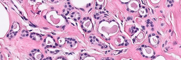
Exemplary floret-like 🌼 tyrosine crystalloids associated with a pleomorphic adenoma #CytoPath #PathTwitter
2
12
43
Collision of good versus bad in this pelvic wash: sheet of benign mesothelial cells versus metastatic high-grade serous carcinoma #CytoPath #PathTwitter
2
19
58