
Mohamed Elsabah
@mohamed_elsabah
Followers
470
Following
1K
Media
109
Statuses
731
Structural echocardiography,cardiology consultant at King Fahad Armed Forces Hospital. Mayo Clinic, Rochester,MN. Mount Sinai Hospital, Univ. of Toronto.
Jeddah, Kingdom of Saudi Arabi
Joined March 2018
RT @iamritu: Sometimes a fibroelastoma on the aortic valve can obstruct a coronary artery #echofirst.
0
16
0
RT @drozgeozden: 🫀 9 Spot Facts Every Cardiologist Should Know About HCM!.📉 Normal EF? ⚠️ Doesn’t always mean normal. 🌀 Apical HCM? 👉 Often….
0
11
0
RT @CASivaram1: #echofirst.✅ Time to conclude, 🙏 for all comments.✅ Diagnosis: ‘Notched' AR on CW Doppler, severe AR (see reference⬇️), sev….
0
6
0
RT @iamritu: #Timing of #Mitral #Regurgitation .Early systole.Late systole .Holosystolic .Biphasic .#color #doppler #echofirst h/t @BonitaE….
0
139
0
RT @EGarciaSayan: 🔥off the press, @ASE360 diastolic Fx guidelines online at @JournalASEcho: 🔺e'≤7 (L) or ≤6 (S), E/….
0
27
0
RT @KFAFHCards: Educational programs w @SCAI_Prez @SrihariNaiduMD .& International Committee 🪑 @mirvatalasnag . Interventional Fellows’ Cou….
0
14
0
These TEEs show right atrial masses identified after catheter insertion.Can you help differentiate which findings are more suggestive of infection versus thrombus?.@ASE360 @KFAFHCards .#echofirst @CASivaram1 @iamritu @purviparwani .@ChamsiPash @OKhaliqueMD @aelsab @MayoClinic
6
12
35
RT @KFAFHCards: Congratulations @KFAFHCards on a great demonstration of IVUS guided PCI of LM with Impella CP support. Operators: @assiri99….
0
7
0

RT @iamritu: 5 Abnormal septum motion patterns on #echofirst that are nonischemic @BijoyKhandheria . . 1.LBBB septa….
0
98
0
57y old mae with severe AR and mild aortopathy , AR jet to LVOT diameter ratio is 60, AI P1/2time is 479ms. How far do you depend on PHT in the assessment of AR?.@KFAFHCards .@iamritu .@hahn_rt .@JaeKOh2 .@EchoSoliman .@ASE360 .@CASivaram1 .@EACVIPresident
8
10
42
RT @m_alfawara: LAA thrombus? 🤔.In AFib patients, a filling defect in the LAA may raise suspicion. A delayed scan (40–60s) can help differe….
0
6
0
42y M. With severe SOB. Diagnosed with massive bilateral pulmonary embolism with large Rt lung infarction. With RV strain underwent catheter embolectomy but still not improved and received fibrinolytic. patient is improving now. @KFAFHCards .@ASE360 .#echofirst.@m_alfawara
1
3
13
RT @ChamsiPash: @mohamed_elsabah @ASE360 @3DEcho360 @iamritu @purviparwani @kfafh Nicely demonstrated. here is what it looks like intraop….
0
3
0
Heart CT was done showing the insertion of the Subaortic membrane near the base of the AV leaflets.Thank you, @m_alfawara. @iamritu.@CASivaram1 .@OKhaliqueMD .@ChamsiPash .@mirvatalasnag .@KFAFHCards
1
4
10
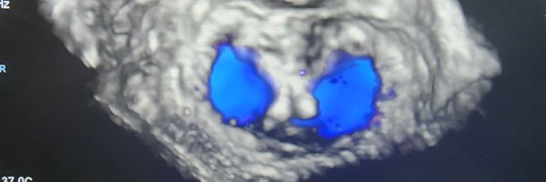
16y M with brother who did cardiac surgical removal of subaortic membrane and AVR at the age of 10y . C/o class IV SOB. found to have a subaortic membrane with severe stenosis & AR.3D demonstrated SA membrane 👌 .#echofirst.@ASE360 .@3DEcho360 .@iamritu.@purviparwani.@kfafh
4
6
35
RT @KFAFHCards: Well done KFAFH Cathlab team.& special thanks to @BSCCardiology for the technical support during the live transmission. @dr….
0
6
0