
Ritu Thamman MD
@iamritu
Followers
32K
Following
214K
Media
7K
Statuses
85K
Associate Professor #cardiologist #SoMe editor @CircOutcomes @JournalASEcho #BOD @ase360 @PittHealthSci @ColumbiaMed @NUFeinbergMed #echofirst #NewJersey girl
University of Pittsburgh SOM
Joined January 2010

Mechanical valve in mitral position: normal appearance. #3DEcho
#echocardiography
#cardiology
#EchoFirst
#CVImaging
#TEE
@ASE360
2
4
12


🫀 A few weeks ago I was puzzled by a child : clear holodiastolic flow reversal in the descending aorta… but Nothing matched. 👇 Acute myocarditis can be accompanied by aortitis, increasing aortic stiffness → diastolic flow reversal #PedsCardio
https://t.co/eyRlpdUVqK
0
2
7

.@BSEcho have launched a new Level 1 library🚀- developed by Jen Gosling, @EchoFoundations, et al, which will support practitioners of emergency echo The first 30 cases are FREE! Find out more: https://t.co/7bKXKHDOEk
3
8
27

#echofirst ✅ Annuloplasty can be done with a band (incomplete circle on echo) or a ring (complete circle) ✅ 3D example of MV annuoplasty band ⬇️ with only residual mild MR & no MS ✅ Marked restriction of PML from annuloplasty @argulian
@iamritu
@purviparwani
@PWesslyMD
2
10
23

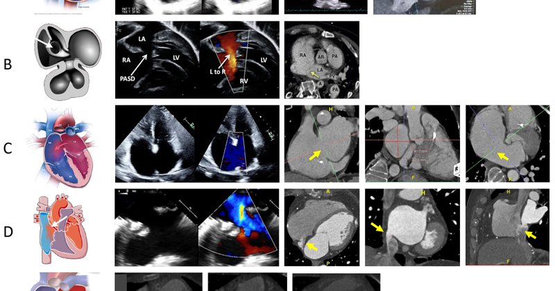
Our article recently published regarding cardiac CTA evaluation of simple intracardiac shunts in adults.@RutgersRWJcards @ppsengupta1 @KamuMaganti @WVUHeart @Heart_SCCT
imrpress.com
Congenital heart disease (CHD) is increasingly detected in cardiac imaging. Effective management of CHD requires thorough imaging of the heart and circulation, extending beyond simple anatomical...
1
10
38
💥A masterclass on Post TAVR PCI 💥What are the Challenges and Technical Considerations in patients undergoing POST_TAVR PCI ⁉️👇 #COMPLEXPCI2025 #TAVR #High_RiskPCI @summitmd_cvrf #1/2

#COMPLEXPCI2025
#HappeningNOW 💥Workshop 5: High-Risk PCI and Calcification 📍Main Arena, Vista Hall 1-2, B2 🕞3:30 PM ~ 4:30 PM * KST (UTC/GMT +9:00) 💥Challenging Case Presentation 5 📍Room 1, Vista Hall 3, B2 🕞3:30 PM ~ 4:30 PM * KST (UTC/GMT +9:00) @summitmd_cvrf
1
24
81
This article was a long time coming!! @PMcardioApp @RobertHermanMD @PendellM Prognostic significance of the discrepancy between STEMI/NSTEMI paradigm and the angiographically confirmed diagnosis of Occlusion Myocardial Infarction: a retrospective cohort study
1
13
47


Strain imaging of LV and RV improves diagnostic sensitivity and predicts arrythmogenic cardiomyopathy (ARC) @ASE360 @pattypellikka @ACCinTouch @iamritu
https://t.co/9aBHAjMFhA
0
3
17

https://t.co/lFISAcAwTO Confirming and quantifying what was suspected - heart failure is evolving. Significant evidence generation implications.
jacc.org
1
18
54

JUST OUT on actual ANATOMIC visualization of the 5-type morphological classification of #TricuspidValve anatomy published previously by @hahn_rt et al, now correlated with #Echo #TEE and schematic by @juliendreyfus1 et al. in #JACCIMG @JACCJournals . A MUST-READ and print-out
0
66
164

Fused BAV (RCC - NCC Phenotype) 20-30% #echofirst 👉BAV-associated aortopathy 👉 Flow Jet right posterior to distal arch = this is type 3 tubular/arch aortopathy 👉 To unveil the mechanism of AR 3DE-based 2DE images are decisive (not the 3D image itself)
5
20
80
Nice #Echofirst trick regarding pericardial effusion and concern for possible continued leak 🎨Use echo contrast! If you see it in the pericardial space= continued leak This case is likely due to a recent PPM lead with continued microperf. @EchofirstB #CardioX #cardiology
1
3
14


It is fascinating that an anatomic study has confirmed what echocardiography previously suggested: tricuspid valve is not tricuspid most of the times due to leaflet scalloping, most commonly of the posterior leaflet. https://t.co/Ed4QZqWcFC
0
2
5

Caseating MAC - I used to see it frequently in Loma Linda with a large aging population..not common in this side of the world Isn’t #YesCCT king though? 👑
6
9
31


-In newborn, Right heart dilation👉🏻TAPVR -Enlarged CS wall may mimic a cortriatriatum sinister. @WGACHDChair @iamritu @escardio @dkthekkoott @CASivaram1 @alexsfelixecho @echoleolopez @DavidWienerMD @alex1708ander @loomba_rohit @swatigar @SIwa23288585 @WithAScalpel @CardioNeo
1
15
54