

Jae K. Oh
@JaeKOh2
Followers
8K
Following
303
Media
187
Statuses
379
Cardiac Hemodynamics, Diastology, Pericardial Diseases, Valvular HD EVOID AS Trial & ECG/Echo AI
Mayo Clinic Rochester
Joined January 2014
Amazing @CASivaram1 electrical and Doppler correlation!.Great to learn something I did not know before.

#echofirst .✅ Time to close, 🙏 for all comments.✅ Answer: Interatrial conduction delay causing a bifid or double atrial reversal (Ar) signal on pulm vein PW Doppler.✅ Evidence for interatrial delay here- #1: P Waves in inferior leads show a terminal neg deflection (late LA


1
5
48
Just finished 5 days of @MayoClinicCV #EchoBoardReview course. We @garvankane provided scholarship to 15 fellows around the country who demonstrated leadership in #Echo education, research, and clinical applications. We are continuing the legacy of @AJamilTajik & #JBSeward.

10
11
109
Congratulations! @EchoSoliman @argulian . #Echo is the Gold Standard for cardiac hemodynamic assessment in clinical practice. We should be able to explain all Doppler velocities and it's patterns in terms of hemodynamics. This excellent timely book helps all of us to do that.

It has finally arrived 🇬🇧! . My latest book, Clinical Applications of Echo Doppler Haemodynamics, co-edited with my colleague @argulian from Mount Sinai, New York, USA, with contributions from my colleague Bernard Bulwer of Brigham and Women’s Hospital, Harvard Medical School,

4
10
85
Diastolic flow reversal due to high RA pressure from myocardial disease occurs with inspiration, restriction. High RA pressure due to RV dysfunction has persistent reversals during diastole. Not all systolic reversals are severe TR which has a delayed peaking.

0
8
52
Happy to see HV Doppler from @argulian . Diastolic flow reversal indicates high RA pressure. Interesting though it happens with expiration. Etiology of high RA pressure? Helpful to see medial e'. See HV Doppler of various etiologies from my Feb 2020 tweet below. @AJamilTajik.

Answer: marked increase in RA pressure. Note high velocity, broad A wave as well as lower velocity, not late peaking systolic reversal.
4
10
50
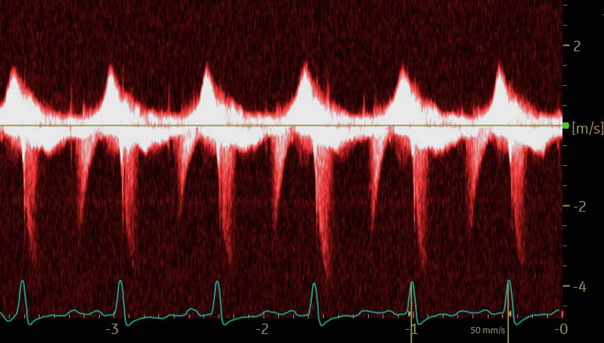
Not sure whether the Doppler velocity below the baseline comes from mitral valve. Those flows occur during isovolumic contraction and relaxation time, typically seen in patients with hypertrophic CM with intracavitary gradient or apical pouch. I have not seen MR doing this.
A patient with shortness of breath. A CW Doppler interrogation across the mitral valve is shown. 1/2
1
4
46
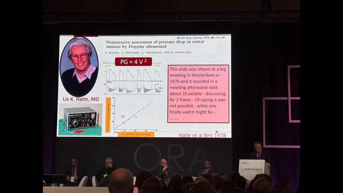
Another simplified EROA is .r*r* x 0.4 when the aliasing velocity is set at 30 cm/ sec. I like this one better since EROA is 0.4 cm2 when PISA radius is 1 cm. Echo Doppler is now the Gold Standard for cardiac hemodynamics in clinical practice. @AJamilTajik #LivHatle.

simplified EROA = (r*r) /2. if we set Va = 0.4 m/s(40 cm/s) and the BP is normal (PG LV-LA = 100 mmHg = Vmax 5 m/s). r*r*6.28*0.4/5. @JaeKOh2.
3
24
99
We describe depth and location of PE. Mild < 1 cm, moderate >1 to 2 cm, and severe >2. But, more important to assess hemodynamics of PE. Amount of PE does not correlate with hemodynamics. Also important to ? effusive constriction which can be assessed .

pubmed.ncbi.nlm.nih.gov
ECP may have unique echo-Doppler features that distinguish it from both CP and tamponade. Our findings suggest that ECP could be diagnosed by echocardiography even prior to pericardiocentesis. ECP...

@JaeKOh2 Please, teach us how to do a correct measure by echocardiography of pericardial effusion. I need to learn asap because I see some incongruencies in the literature! Thanks!.
1
13
64
I agree with you @bobmiles71 We need to make interpretation of DF assessment simple as possible. At @MayoClinicCV, we report grading and it's associated filling pressure as normal , mild to moderately elevated (gr 2), or severely elevated (gr 3).

@JaeKOh2 @SFNagueh @ASE360 @AJamilTajik Thanks. BTW- We use up to date echo criteria for evaluating diastolic dysfunction, however, in our final reports we referred to LV diastolic dysfunction as either “mild, moderate or severe”. We find this helps our referring doctors and the patients comprehend better.
1
1
17
Hopefully, 2025 DF guideline @SFNagueh will minimize indeterminate DF based on fewer parameters. Adjudication of diastolic dysfunction based on clinical history should be avoided. You will find our @ASE360 Editorial interesting. @AJamilTajik.

Interesting results 😳.Prof. @JaeKOh2 🌟.
5
19
63
Hemodynamic cath was done to confirm constriction shown by Echo Doppler because the patient did not have JVP distention and IVC did collapse. LVEDP > RVEDP at rest. Both increased with exercise c/w RCM or CP. CT showed calcified pericardium over the LV. Example of Left-sided CP


The topic was "Pitfalls of hemodynamic assessment" and I showed challenging cases by cath hemodynamics. What is the condition shown in a HF patient by rest and exercise hemodynamics shown? Happy Holidays!

2
28
117
Happy New Year from Rochester, Minnesota!


#HappyNewYear2025 .#BELIEVE #BELIEVE #BELIEVE #BELIEVE.Wishing everyone a healthy happy 2025 #CardioTwitter .@mswami001 @mmamas1973 @DrMarthaGulati @purviparwani @AHagendorff @rajdoc2005 @pattypellikka @JudyHungMD @RigolinVera @KTamirisaMD @JaeKOh2 @DrJenniferCo_Vu @LucySafi

2
1
38
The topic was "Pitfalls of hemodynamic assessment" and I showed challenging cases by cath hemodynamics. What is the condition shown in a HF patient by rest and exercise hemodynamics shown? Happy Holidays!

#echofirst that was a cool session .#EuroEcho2024 .@JaeKOh2 .@echo_batman .@iamritu .@NMerke .@ASE360 .@EACVIPresident


6
18
78
I apologize for delayed response > a year later, but being late is better than never. @ecocardio_cl HV.#colorMmode is a beautiful example for timing of cardiac events. In someone with HF symptoms, the color M-mode alone is good enough for Dx of CP. Agree & Happy Holidays!.


1
17
77
RT @MayoClinic: At Mayo Clinic, we honor the spirit of Christmas and the message of hope, love and compassion it represents. Whether you're….
0
13
0
RT @KrisBergHansen: In patients with #cardiogenicshock 🫀worsening global longitudinal #strain #GLS associates with poor outcomes and surpas….
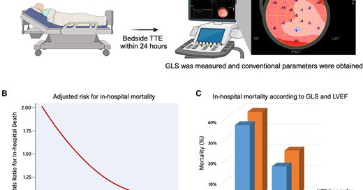
academic.oup.com
AbstractAims. Cardiogenic shock (CS) is a critical manifestation of severe cardiac dysfunction, necessitating precise evaluation of left ventricular (LV) f
0
4
0
RT @ChamsiPash: Stay tuned tomorrow, will have @JaeKOh2 presenting to us @MethodistHosp about constrictive pericarditis, timely after the p….
0
31
0
What an amazing skill and result of apical myectomy for #apicalHCM by @HSchaffMD ! Meticulously avoiding injury to papillary muscle. Presented at our annual #EchoNY. The procedure improves LV cavity size, LV filling pressure , and clinical outcome. @MayoClinicCVS @MayoClinicCV
4
36
119
This is annual #EchoNY by @MayoClinicCV & @hahn_rt .We just completed a heated session on transcatheter vs surgical intervention for VHD with @HSchaffMD @MayraGuerreroMD @OKhaliqueMD #SariPadang. Passionate discussion by ALL!


1
6
43