

Dr. Roig
@doctor_roig
Followers
4,930
Following
452
Media
300
Statuses
4,070
Author of @ekgdx | Co-Founder of Roinova | EKGs lover | Futurist | Innovator | Revolutionizing EKG learning | #AI | #ekgdx
🇺🇸 🇨🇺
Joined May 2012
Don't wanna be here?
Send us removal request.
Explore trending content on Musk Viewer
Robert Fico
• 168029 Tweets
Biden and Trump
• 122502 Tweets
Slovakia
• 108426 Tweets
Peru
• 97713 Tweets
Wicked
• 75671 Tweets
MY LOVE IS KING KONG
• 66937 Tweets
サーティワン
• 50133 Tweets
キダ・タローさん
• 46952 Tweets
Samurai
• 44092 Tweets
Assassin's Creed
• 38679 Tweets
Eslovaquia
• 26636 Tweets
浪花のモーツァルト
• 25401 Tweets
スロバキア
• 24037 Tweets
Ubisoft
• 23816 Tweets
Vargas
• 23589 Tweets
Yasuke
• 21866 Tweets
Scrap
• 18378 Tweets
Ellie
• 17700 Tweets
Ludmilla
• 17379 Tweets
Glinda
• 14422 Tweets
Daniel Caesar
• 13388 Tweets
Valhalla
• 12555 Tweets
チンチロ
• 12482 Tweets
Ivete
• 12272 Tweets
最高顧問
• 11076 Tweets
Nitro
• 10166 Tweets


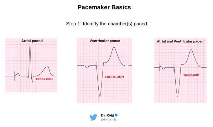
1/ Today's 🧵 is about the
#pacemaker
basics.
The objective of this post is to clarify several doubts in a simple way.
#CardioTwitter
I will explain it with simple steps and classic examples so you can understand better.
@ekgdx
8
342
1K
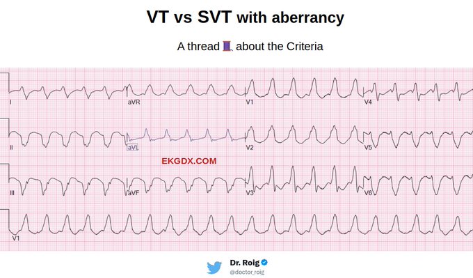
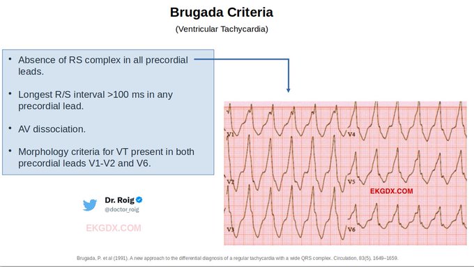
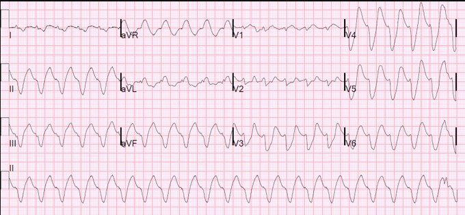
1/ Today's 🧵is about "VT" versus "SVT with aberrancy".
The aim of this thread is to provide basic tips on how to apply some of the most used criteria that might be helpful in diagnosing VT.
#CardioTwitter
Note that the following features are suggestive of VT, but their absence
14
374
1K
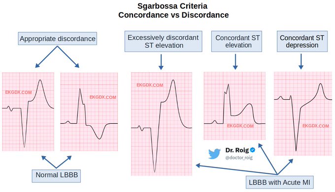
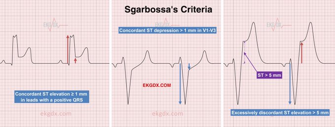
1/ Today's 🧵 is about the
#Sgarbossa
Criteria and
#Concordance
vs
#Discordance
in simple words.
The objective of this post is to clarify any doubts in a simple, graphic and didactic way.
#CardioTwitter
@ekgdx
12
245
792
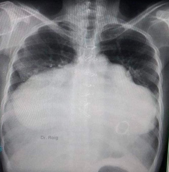
This is the biggest heart I've ever seen.
40 years old 👩🦰 with a history of dilated heart disease (no more info). She sent me this pic via DM.
#CardioTwitter
34
76
542
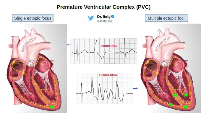
1/ Today's 🧵 is about the premature ventricular complex (
#PVC
).
The objective of this post is to refresh basic concepts. I will explain it in a simple way with classic examples that may help you.
#CardioTwitter
@ekgdx
1
149
539
1/2
The "frog sign" is nothing more than the cannon-type "a" waves produced by the nearly simultaneous contractions of the atria and ventricles while the mitral and tricuspid valves are closed in the setting of a rapid tachyarrhythmia.
🎥
@fazalabul
edited by 🙋♂️
Short🧵
3
117
490
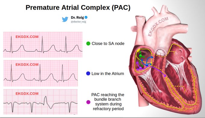
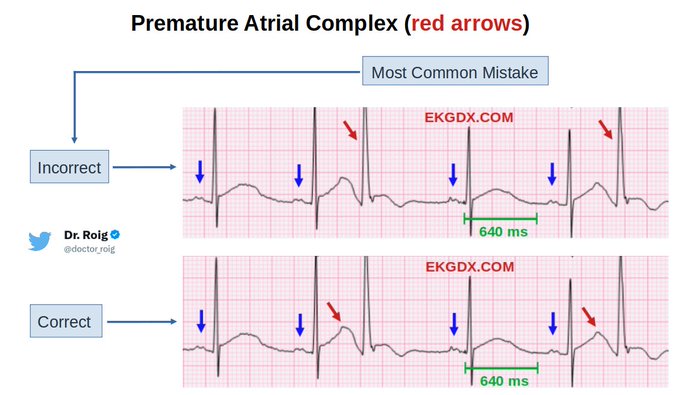
1/ Today's 🧵is about "Premature Atrial Complex (PAC)".
The aim of this thread is to refresh basic concepts that may help you to identify the different types of PACs.
I will explain it in a simple way with classic examples.
#CardioTwitter
#ekgdx
@ekgdx
6
116
471
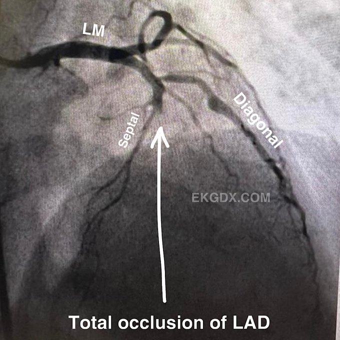
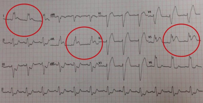
1/ The importance of serial
#EKG
in acute myocardial infarction.
An interesting case with a series of three EKGs with
#echofirst
and
#angiogram
as well.
#CardioTwitter
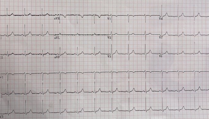
A 52-year-old woman, smoker with chest pain for one hour.
EKG
#1
was performed in triage 🧵👇

12
85
331
@EM_RESUS
1- Endotracheal tube.
2- The absence of chest tubes/central lines indicates old post op (sternal wires), most likely CABG bc no artificial valves.
3- Single chamber pacemaker
4- Deep sulcus sign indicating left pneumothorax (due to CPR??).
5- Cracked ribs (due to CPR??).
6-
11
3
222
1/ Today's 🧵is about EKG Challenges.
It is dedicated to those who need to take their
#ECG
skills to the next level and all
#cardiology
fellows in training.
#CardioTwitter
@ekgdx

4
51
220
1/ Official announcement.
Today is
#WorldHeartDay
.
In honor of such an important date, I am thrilled to announce my little contribution to the EKG world.
Thread 🧵
#CardioTwitter
Here we go👇

8
43
164
@Lap_surgeon
Looks like the stomach (very distended with air) is in the left thoracic cavity, causing a tension displacement of the mediastinum, in the setting of a diaphragmatic rupture, which is suggestive of a gastrothorax tension. Hemodynamic compromise is imminent.
3
5
158
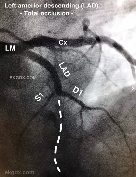
The case of the day (yesterday).
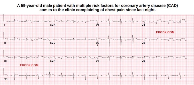
61-year-old 👨🏻🦳 with chest pain 🚨🚨.
#cardiotwitter
where is the culprit lesion 👀?
I’ll post the angiogram 🎥 in few hours.
#ekgdx
#RadialFirst
#STEMI
#MedTwitter
#ecg
#ekg
#culprit
25
28
129
@AvrahamCooperMD
@AvrahamCooperMD
similar case 👀
This is the biggest heart I've ever seen.
40 years old 👩🦰 with a history of dilated heart disease (no more info). She sent me this pic via DM.
#CardioTwitter
34
76
542
1
14
122
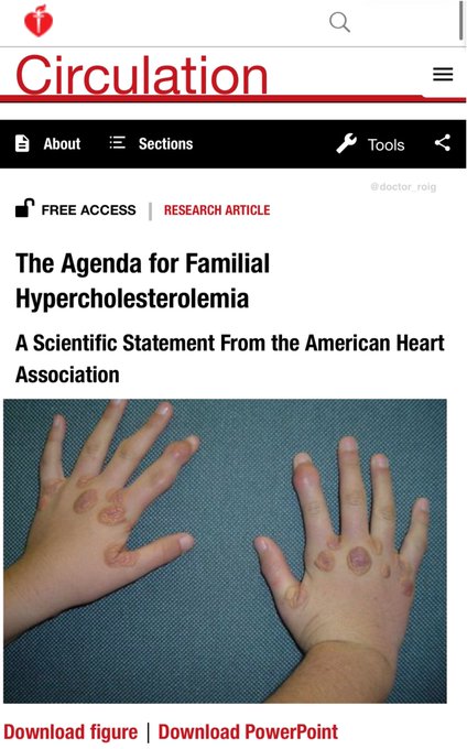
@IhabFathiSulima
This is a classic sample of Familial Hypercholesterolemia.
Thanks for sharing
@IhabFathiSulima
#ekgdx
4
8
97
A normal EKG does not mean that the patient is not having acute ischemia, injury, or infarction.
🚨 47 yo male with chest pain for a few days.
Angio below 😳
#CardioTwitter
#RadialFirst
18
28
90
I have the privilege of announcing that my online EKG book is already available.
Open your Laptop and 👉
Check this out
@smithECGBlog
@Dr_DanMD
@Vadeboncoeur_Al
@GARCIAEDINSON95
@yourheartdoc1
@DocNikko
@narrowQRS
@mmamas1973
@EPeeps_Bot
@ShariqShamimMD
8
30
85
STEMI Alert 🚨 # 3 of the day. 69 y/o Male c/o Chest Pain and SOB. Cath pictures below 👀. Take a look
@smithECGBlog
@EM_RESUS
@Vadeboncoeur_Al
13
27
87
@IhabFathiSulima
This is classic sample of Thoracomelia, witch consist in the presence of extra-limb attached to the thorax. In general, Polymelia is a congenital anomaly, which is defined as the presence of accessory limbs attached to various body regions.
2
6
72
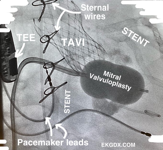
Case of the day.!!!
#CardioTwitter
does he need more ??
You don’t get this picture very often. Saved ✅.
#RadialFirst
#echofirst
#TAVI
#valvuloplasty
#mitral
#medicine
#ekgdx
6
19
70
I wake up and see this beautiful EKG in my text messages. Patient has severe chest pain🚨.
A friend of mine asking for my opinion.
#cardiotwitter
what is happening here?
15
15
69
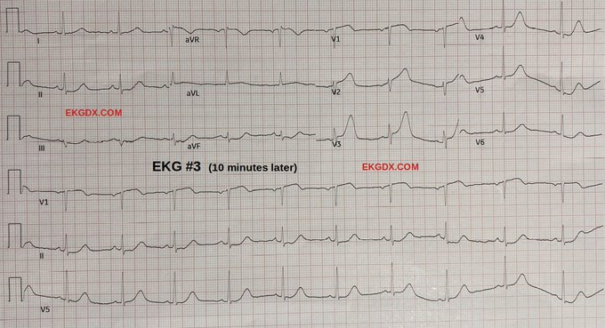
10/ End
Learning point:
Performing EKG every 10 minutes in patients with suspected acute MI can save many lives and avoid delays in appropriate management.
Thanks for reading this teaching case.
Here I share some classic EKGs that you might like
4
10
67
5
12
61
1/ The importance of
#echofirst
in acute
#myocardial
#infarction
.
An interesting case with LV
#thrombus
in the setting of anterior acute MI with echo and
#angiogram
included.
#CardioTwitter
🧵
2
19
55
1/ Let’s talk about Concealed Conduction
#CardioTwitter
.
Manifestation of
#Concealed
#Conduction
are numerous because the variations of the site impulse formation, the different effects of the anatomical site, the direction of the concealment, and the changing
3
14
46
@daguitovaldes
Si somos objetivos, la ayuda arbitral empaña todo el debate en cuanto a resultados (por desgracia). Por tal motivo, los blancos salen en ventaja (teniendo en cuenta tan importante aspecto). Saludón hermano
@daguitovaldes
8
1
44
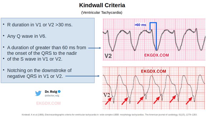
3/ In the past 40 years, several algorithms or criteria have been developed to differentiate VT from SVT. Here I show you some of them:
Kindwall Criteria
Article:
Courtesy of
1
5
43
1/2
Delta wave (Dw) and relationship to location of the accessory pathway.
If Dw + in V1, aVF and - in aVL= L lateral.
If Dw + in V1, aVL and - aVF = L post./septal.
If Dw + in aVL and - V1, aVF = R post./septal.
If Dw + in aVF, aVL and - V1 = R lateral/anterior
2
9
40
14/ Wide QRS Mimic
Here is a sample of "Wide QRS Mimic" due to severe ST elevation in the setting of pLAD occlusion.
Courtesy of
@narrowQRS
2
8
41
@EPWaveDoc
Nice catch
@EPWaveDoc
1/2
The "frog sign" is nothing more than the cannon-type "a" waves produced by the nearly simultaneous contractions of the atria and ventricles while the mitral and tricuspid valves are closed in the setting of a rapid tachyarrhythmia.
🎥
@fazalabul
edited by 🙋♂️
Short🧵
3
117
490
2
4
38
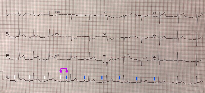
STEMI case last night (culprit was prox RCA).
BUT!!!!
What’s happening here (purple arrow)? Your opinion.
White and blue (P waves).
@narrowQRS
@SergioPinski
@Dr_OkeefeECG
@jvillacastin
@bordistef
@Hapa_EP
@Dr_Nazarian_EP
@DrJasonAndrade
#Epees
#cardiotwitter
6
6
38
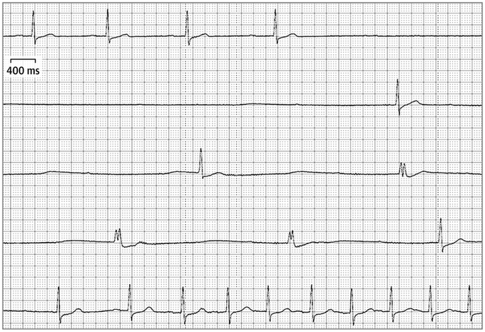
How many times have you seen only P waves on the monitor ?
#Epees
#cardiology
#medicine
#ekgdx
#cardiotwitter
#only_p_waves
#3AVB
4
9
35

Finally, the FDA recommends that Hydroxychloroquine no longer be used for the treatment of
#COVID19
.
Once again, the evidence-based science proves the right thing.
The scientific method should never be questioned by the simple whim and ignorance of anyone.
3
6
34
How many times have you seen AFIB and Sinus rhythm at the same time?
#cardiotwitter
#EPeeps
#cardiology
#ekgdx
2
6
34
2/ The Sgarbossa criteria were initially introduced over two decades ago to enhance the diagnostic precision for MI in the setting of LBBB. This criteria is widely accepted as one of the most valuable tools to assist in the diagnosis of MI when LBBB is present.
Here you can see
1
12
35
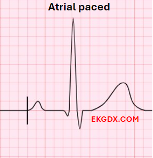
2/ Step 1: Identify the chamber(s) paced.
Let's look at the classic examples.
A spike occurring before the P wave usually indicates atrial pacing.
1
7
34
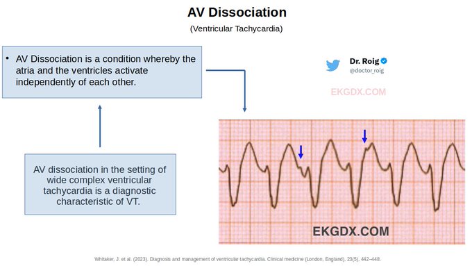
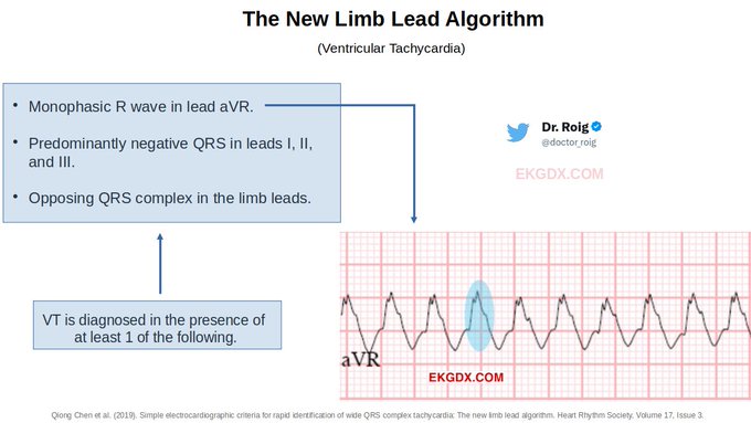
7/ After having analyzed some criteria, let's look at some electrocardiographic characteristics that suggest VT.
AV dissociation: Although the presence of AV dissociation is highly suggestive of VT, its absence does not exclude VT. A single dissociated P wave at the onset of
1
3
34
1/
#cardiotwitter
thanks for the debate.
The culprit was the prox RCA (👀🎥)
It's a bit tricky bc it gets confused with the LAD as the culprit.
This pattern have been described and usually associated with the RCA as culprit. Read the following papers in the next tweet👇.
2
1
30
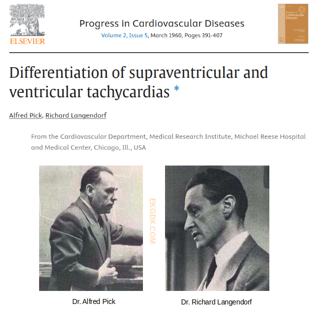
2/ History
In 1960 Dr. Alfred Pick and Richard Langendorf published, “Differentiation of supraventricular and ventricular tachycardia.” Sixty years later, differential diagnosis of wide QRS tachycardia on the electrocardiogram remains a challenging exercise.
Article:
2
2
32
@docramiro
Another similar case!!!
This is the biggest heart I've ever seen.
40 years old 👩🦰 with a history of dilated heart disease (no more info). She sent me this pic via DM.
#CardioTwitter
34
76
542
0
0
31
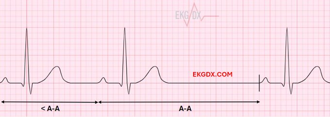
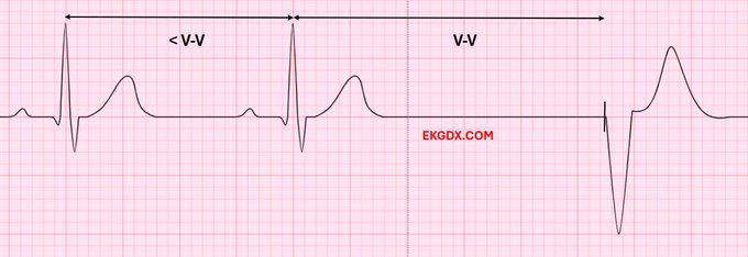
8/ Step 3: Identify the chamber(s) sensed.
Atrial pacemaker: Adequate atrial sensing is confirmed when intrinsic atrial activation (native P wave) is followed by either (1) a native P wave occurring at an interval shorter than the A-A interval or (2) an atrial-paced beat that
2
7
31
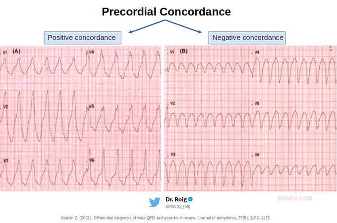
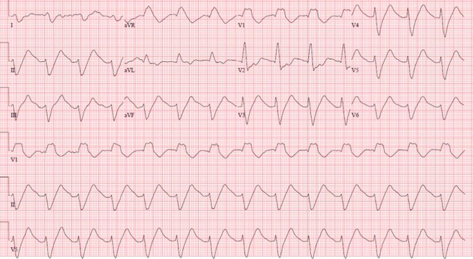
10/ Precordial Concordance
During precordial concordance, there is no R/S transition and is suggestive of VT (PPV 90%–100%, sensitivity 88% [Miller JM. et al 2006]). Although precordial concordance is highly suggestive of VT, its absence does not exclude the diagnosis.
1
4
29
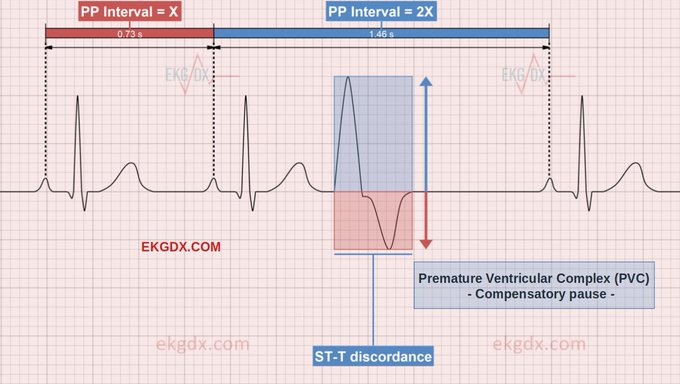
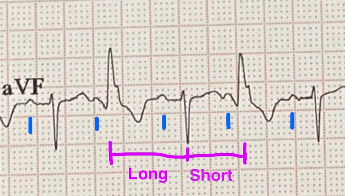
3/ In regards to the compensatory pause:
Complete pause: The premature beat does not reset the sinus pacemaker due to either VA block or the sinus node being refractory at time of premature impulse arrival. The subsequent sinus beat occurs on time based on the underlying sinus
1
6
28
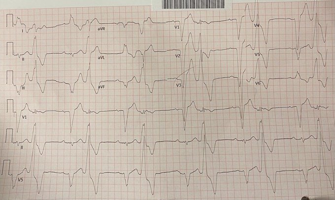
13/ Severe hyperkalemia. Courtesy of
@smithECGBlog
The patient had a K of 8.1 mEq/L and a very low ionized Calcium.
1
5
29
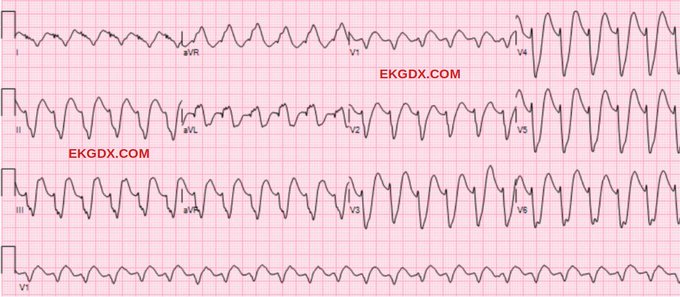
12/ VT Mimics
It is important to clarify that on many occasions we will face ECGs that may look like VT but are not, and are the so-called "VT Mimics". Let's look at some examples:
Flecainide overdose. Courtesy of
@SergioPinski
1
2
28
@Sthanu5
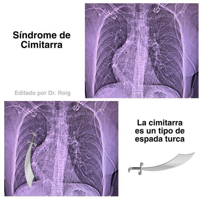
The malformation was first described in 1836 by Cooper and Chassinat, however, the scimitar sign was described by Halasz et al in 1956. They described the anomalous pulmonary vein showing a vertical course which takes the form of a scimitar (a type of Turkish sword).
0
8
27
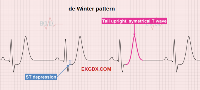
2/ The de Winter pattern holds significance as it is linked to the occlusion of the proximal left anterior descending coronary artery (LAD) when identified in the electrocardiogram (ECG) of individuals experiencing chest pain or displaying a history suggestive of acute coronary
1
8
28
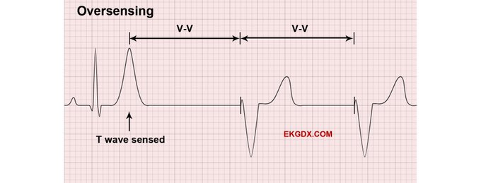
11/ Oversensing is identified when, according to timing intervals, pacing spikes that should have been initiated after a native P wave or QRS complex were not. This leads to a paced beat that appears later than expected. In the case of ventricular pacing, oversensing occurs when
2
7
28
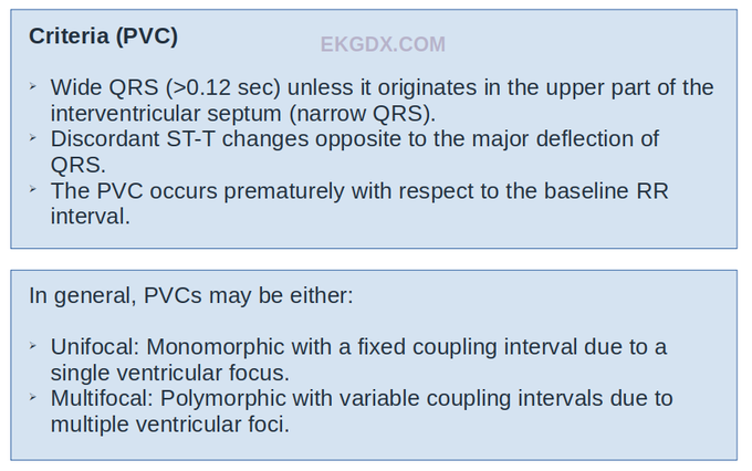
2/ Criteria
Premature ventricular complex occurs when a premature beat arises from an ectopic focus within the ventricles. In the majority of cases, PVCs have no known cause and may occur spontaneously.
1
5
28
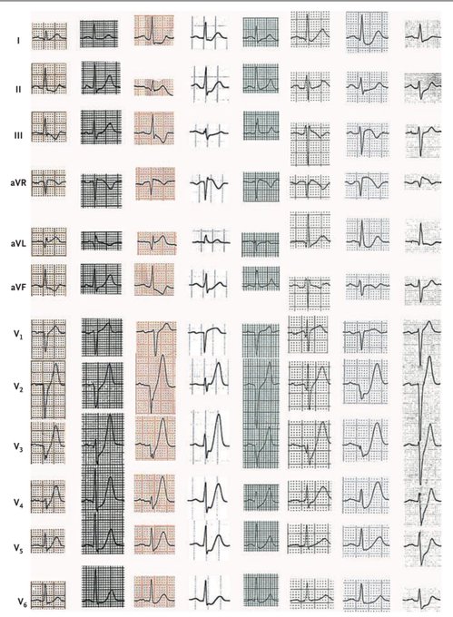
A professor of Pennsylvania ask me: Your software is capable to generate almost any EKG ? Send me this pattern:
EKG with concave STE in I and aVL
HR: 120 x min
Inverted U wave in I and aVL
Inverted P wave in aVL
Notched P wave in I
Full EKG 12 leads.
Me: I sent the proof
5
9
27
How many time have you seen two TAVI in the same picture?
Case of the day !!!
Save it ✅
#cardiotwitter
#ekgfirst
#ekgdx
#TAVI
#TAVR
#MedTwitter
4
5
25
@freddier
Esos “ingenieros” sufren de algunos males, dentro de los cuáles, el NO darse cuenta (x su EGO) a que TODO trabajo (sea CRUD o no) tiene IMPORTANCIA y que indiscutiblemente, les duele NO ser parte de esas GRANDES empresas que mencionas ..
1
1
26
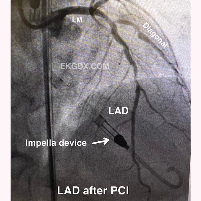
The
#angiogram
shows 99% stenosis of the LAD.
See the EKG above 👀👆.
#MedTwitter
#pci
#ekgdx
#CardioTwitter
#LAD
#stenosis
3
2
25
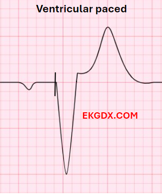
3/ A spike occurring before the QRS complex typically indicates ventricular pacing.
1
8
26
3/ History
Dr. Robbert Jan de Winter is credited with initially describing it in 2008 in a letter to the editor of the New England Journal of Medicine. What Dr. de Winter characterized was “1- to 3-mm upsloping ST-segment depression at the J point in leads V1 to V6 that
2
7
27
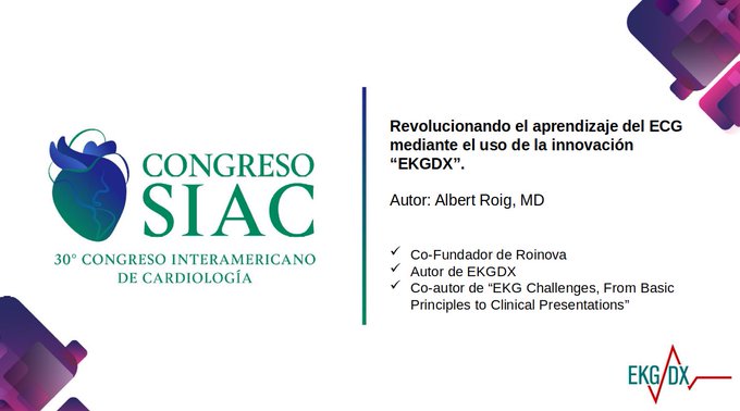
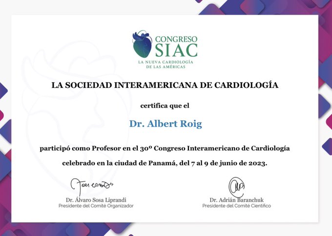
1/ En el día de ayer, tuve el privilegio de presentar nuestra innovación EKGDX en el Congreso de Cardiología SIAC 2023.
Por tal motivo, les comparto algunos aspectos importantes de la presentación
Abro hilo🧵
@SIAC_cardio
@ekgdx
1
6
25
12/ Step 4: Identify pacemaker malfunction.
Sensing abnormalities: Undersensing occurs when, based on timing intervals, pacing spikes that should have been inhibited by a native P wave or QRS complex were not. This leads to a paced beat that appears earlier than expected.
For
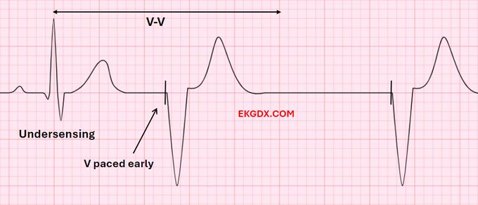
1
8
26
16/ END
Thanks for reading this 🧵.
If you find this content helpful, please:
1- Follow
@doctor_roig
and
@ekgdx
for more.
2- Re-post the first tweet for support.
3- Activate my bell and don't miss my next thread.
3
5
26
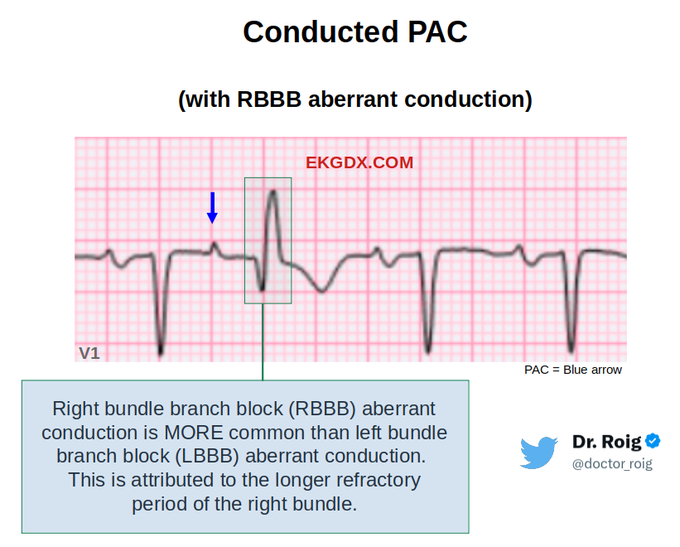
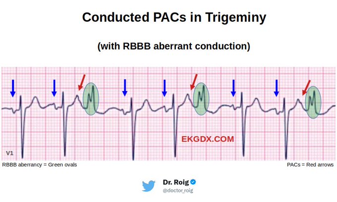
2/ PAC occurs when an ectopic focus within the atria, generates an action potential before the next scheduled sinus beat.
In general:
*PAC can be Conducted normally, Aberrantly conducted or Blocked (non-conducted).
*PAC can be conducted with short PR, normal PR or long PR
1
6
26
LAD coming from the RCA.
Anatomical variations sometimes are present.
What a nice view..!
#cardiotwitter
#cathlab
#RadialFirst
#echofirst
#LAD
#RCA
#cases
#medicine
#rare
#ekgdx
#Cardiology
#fellows
#interventional
0
9
23
3/ The Sgarbossa Criteria were initially introduced in 1996 by
@ElenaSgarbossa
et al. They used data from the famous GUSTO-1 trial.
Article🔗
1
3
25
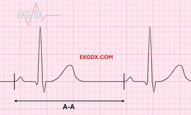
5/ Step 2: Identify the timing intervals from two consecutively paced beats.
Here a sample of A-A interval in the setting of atrial pacing.
1
7
25
2/2
This causes an abrupt increase in right atrial pressure that is transmitted in reversal of flow in the superior vena cava resulting in jugular venous pulsations.
#MedTwitter
#CardioTwitter
#frogsign
#Cardiology
#medicine
#media
1
4
25
11/ I think it's time to apply what we've learned:
1: What criteria are present in this EKG?
2: This is VT?
3: SVT with aberrancy?
3
2
25
64 yo male w chest pain. There is severe LM lesion. EKG (below) shows subtle STE in aVR, ST-T changes suggesting myocardial ischemia in V1-V4, RBBB pattern and fQRS in inferior leads suggesting scar from old MI (abnormal q waves).
#Cardiology
#RadialFirst
#echofirst
#ekgdx
7
7
23
9/ Ventricular pacemaker: For proper ventricular sensing: It is confirmed when intrinsic ventricular activation (native QRS complex) is consistently followed by either (1) a native QRS complex occurring at an interval shorter than the V-V interval or (2) a ventricular-paced beat
1
6
24
Learning EKG is still a problem for many. It's time to change the way we learn it? 🤔.
A new generation.
A new method.
A new era.
See you in Panama 🇵🇦.
@SIAC_cardio
#congress
#siac

1
6
23
@smithECGBlog
That’s sad 😞. Make me upset any time I see an interventionalist refuse to put the patient on the table (cath lab) because the EKG do not meet the STEMI criteria 🤦♂️. Guidelines and just Guidelines. Our profession is more than that.
0
0
23
This is one of the reasons why it is better to use Brilinta as a load and not as a long-term treatment. Pharmacology (is our weapons in this war)

0
9
22
@EcgOxford
@MaruanCarlos
@The_Nanashi_O
@DruvBhagavan
@UlhasDr
@ECGfan
@DidlakeDW
@ecgrhythms
@syamkumarmd
@rajivasr
@FloydECGs
@DrRajeshG1
@CarlosVergaraMD
@DocNikko
@juliogilpereira
@Vadeboncoeur_Al
@TahaMD_EM
@drhbkmd
1/2 Sinus tachycardia (blue) with frequent premature QRS complexes with RBBB pattern occurring in bigeminy which could either be supraventricular with aberrant conduction or PVC. On careful analysis, note that the PP intervals are regular. In contrast, the RR intervals …
2
2
22
13/ References
Here a link with several articles about pacemaker.
1
9
23
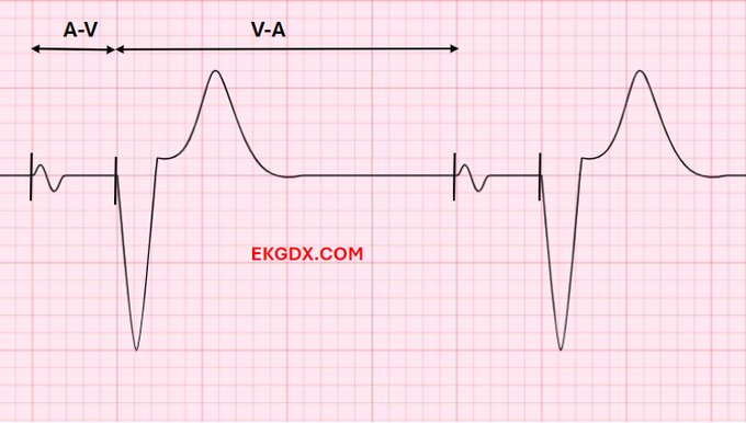
7/ Here a sample of A-V and V-A intervals in the setting of dual-chamber pacing.
1
6
22
A nightmare !!
#Cardiotwitter
🎥 Sent by a friend without the history.
In case you are having a bad day, others have it worse.
#ekgdx
5
4
19
2
3
22
Official announcement ✅
It is an enormous privilege for me to work together with Dr. Alfonso Tolentino in the development of Volume II of our educational platform on electrocardiography.
#cardiotwitter
3
3
19
15/ If you want to practice with EKG Challenges, check this out
1/ Today's 🧵is about EKG Challenges.
It is dedicated to those who need to take their
#ECG
skills to the next level and all
#cardiology
fellows in training.
#CardioTwitter
@ekgdx
4
51
220
1
1
21
6/ PVCs may occurs in a pattern of:
Isolated: only one PVC (see sample below).
Bigeminy: every other beat is a PVC.
Trigeminy: every third beat is a PVC.
Quadrigeminy: every fourth beat is a PVC.
Quintageminy: every fifth beat is a PVC.
Couplet: two consecutive PVCs.
Ventricular
1
4
21
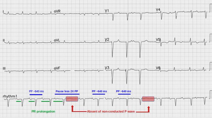
12/ Failure to capture (Atrium or Ventricle) occurs when a pacing spike is released but fails to stumilate the atrium or ventricles. In other words, pacing spikes are not followed by a P wave or QRS complex.
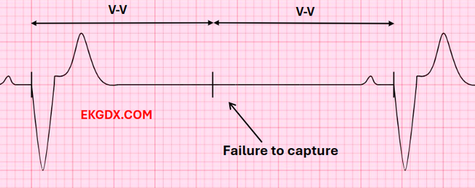
1
5
20
@daguitovaldes
Mi opinión:
Si Bellingham está haciendo números al nivel de Cristiano, es mucho decir !!!
En este momento, está en el top 3 mundial.
0
0
16
I'm already in Panama 🇵🇦🙋♂️.
See you at the Interamerican Congress of Cardiology to talk about EKG learning through the use of innovation.
@SIAC_cardio
@adribaran
#ekgdx
1
4
20
@AndreMansoor
These observations are indicative of severe tricuspid regurgitation. The side-to-side head bobbing is a consequence of the impact caused by the bolus of regurgitant blood moving upward in the neck and consequently visible on the scalp, leading to the manifestation of Lancisi's
0
0
18
2
2
19
@ShariqShamimMD
@EM_RESUS
@smithECGBlog
@ECGfan
@DrMarthaGulati
@mirvatalasnag
@mmamas1973
@iamritu
@yourheartdoc1
@DrRyanPDaly
@cardiojaydoc02
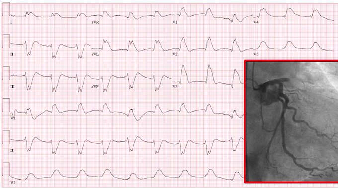
The most common cause of global T-wave inversion is myocardial ischemia, which almost always shows a reciprocal upright T wave in lead aVR. When T waves are positive in aVR, the patient is at increased risk of death. Is hard to say the culprit, but I go with LM vs Prox LAD.
1
1
19
Con mucho cariño lo guardaré por sobradas razones.
Fue un Congreso inolvidable para mí.
Gracias
@SIAC_cardio
Gracias
@adribaran
1
5
19
9/ Another sample of acute MI in the setting of LBBB.
Red circle shows concordant ST segment elevation. Courtesy of Dr. Carlos Pineda from ECG Weekly.
1
2
19
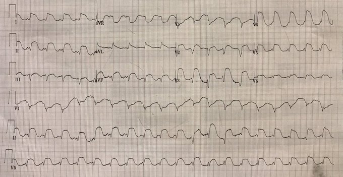
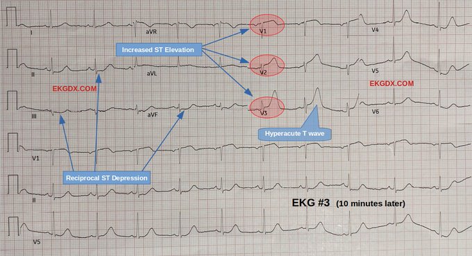
3/
10 min later, third EKG was performed.
Fentanyl (50 mcg) IV given with no pain release.
The patient begins with signs of facial sweating.
Vital signs remain stable.
1
3
20