
Gang Wen
@GangWen1
Followers
43
Following
301
Media
1
Statuses
53
Chemical biology, expansion microscopy, and probe design.
Joined July 2019
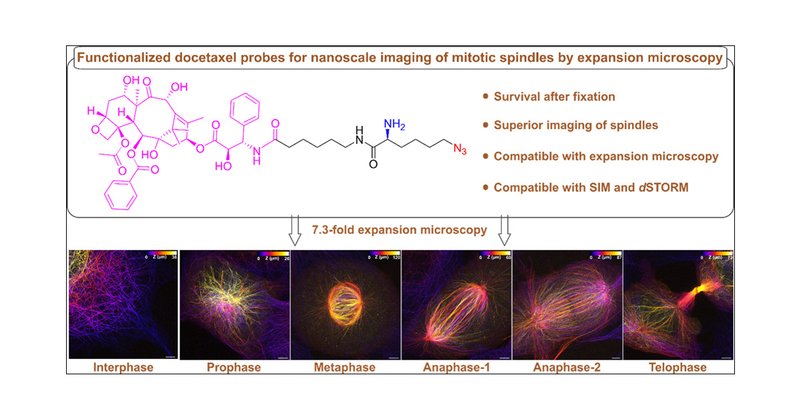
Our new functionalized docetaxel probe enables superior visualization of dense mitotic spindle structures during cell division by expansion microscopy! | Journal of the American Chemical Society
pubs.acs.org
Visualizing the ultrastructure of mitotic spindles, the macromolecular machines that segregate chromosomes during mitosis, by fluorescence imaging remains challenging. Here we introduce an azido- and...
1
4
17

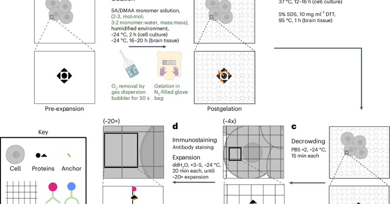
The new technique is described in a collaborative paper that published today @naturemethods
https://t.co/ZjzwwuW18M
@eboyden3 @ChemicalBiology @broadinstitute @kochinstitute @HHMINEWS @ChemistryMIT @mitbrainandcog @MITdeptofBE @ScienceMIT @MITEngineering
nature.com
Nature Methods - 20ExM achieves isotropic ~20× expansion of cells and tissues in a single shot for super-resolution imaging with <20-nm resolution on a conventional microscope.
34
9
13

Getting rid of linkage errors and labeling protein of interest directly at the desired site via click chemistry. Gerti Beliu @unnaturalGerti talks click labeling for improving fluorescence microscopy and imaging substantially. Check out his works here https://t.co/uMXsHt8Iic
1
4
22
Fabulous Dominic @DominicHelm now takes the stage to tell us about Picorulers as a prominent calibration ruler in superres imaging #FOM2024
https://t.co/vyk3b6yfIU
0
3
14

NEW📰🚨:"Protein-based PicoRulers set new standards for the verification of sub-10 nm super-resolution microscopy methods under cellular conditions." ▶️ https://t.co/PaEsavCpYZ
1
10
52

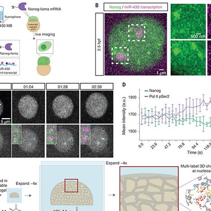
Our work describing Chromatin Expansion Microscopy is now out in @ScienceMagazine
https://t.co/ysL8Q1xmqA Check out how we were able to visualize transcription, chromatin, and TFs with molecular resolution in developing zebrafish embryos to understand gene regulation!
science.org
In vivo imaging with chromatin expansion microscopy reveals principles of pioneer factor–controlled transcription in zebrafish embryos.
15
113
371

How do you image the large and the small at the same time? We developed new 🔬 technology to image centimeter-scale specimens - including whole mouse brains 🧠 - with diffraction-limited resolution and without sectioning. #mesoscale #imaging
https://t.co/Kl7ZURNpdM 🧵 (1/n)
30
196
617

Do you want to label a protein (for fluorescence detection, FRET, EPR, etc.) but don’t know what residues are most suitable? Check out our “Labelizer” pipeline to predict the best labeling sites! Great collaboration with Thorben Cordes @PSB_lab
https://t.co/X4DLscTbxz 🧵 (1/n)
3
53
233
Visualizing actin filament with high quality in ExM is difficult, even with original trifunctional phalloidin linkers, due to the low phalloidin density on the surface of actin monomers (One phalloidin/actin monomer). Detailed optimization is here.

biorxiv.org
Expansion Microscopy (ExM) revolutionized the field of super-resolution microscopy by allowing for subdiffraction resolution fluorescence imaging on standard fluorescence microscopes. However, it has...
1
1
10

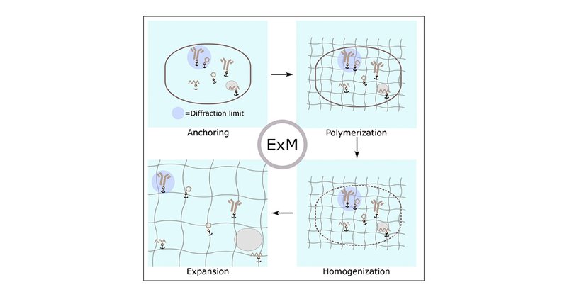
🔔 Everything you ever wanted to know about expansion microscopy and then some: https://t.co/dHS6Y2N41p 🔬↔️↕️
pubs.acs.org
Expansion microscopy (ExM) is a newly developed super-resolution technique, allowing visualization of biological targets at nanoscale resolution on conventional fluorescence microscopes. Since its...
0
8
34
So humbled and glad to have it published. It has been one and half years since I started to write it, the first chapter of my Ph.D thesis. Hope that you could get some ideas of ExM from chemical aspects.
pubs.acs.org
Expansion microscopy (ExM) is a newly developed super-resolution technique, allowing visualization of biological targets at nanoscale resolution on conventional fluorescence microscopes. Since its...
0
2
8
Interesting
We present Compression #Microscopy (CoMic) as a new #superres modality. By compressing the sample it is made inaccessible by conventional imaging, thus necessitating superres. CoMic allow imaging of macro-structures by superres, and could make diffraction-limited imaging obsolete
0
0
1


25
152
2K
We are excited to share our recent work. A Universal Labeling Strategy for Nucleic Acids in Expansion Microscopy
pubs.acs.org
Expansion microscopy (ExM) enables the nanoscale imaging of ribonucleic acids (RNAs) on a conventional fluorescence microscope, providing information on the intricate patterns of gene expression at...
1
7
16
Evaluation of Direct Grafting Strategies via Trivalent Anchoring for Enabling Lipid Membrane and Cytoskeleton Staining in Expansion Microscopy
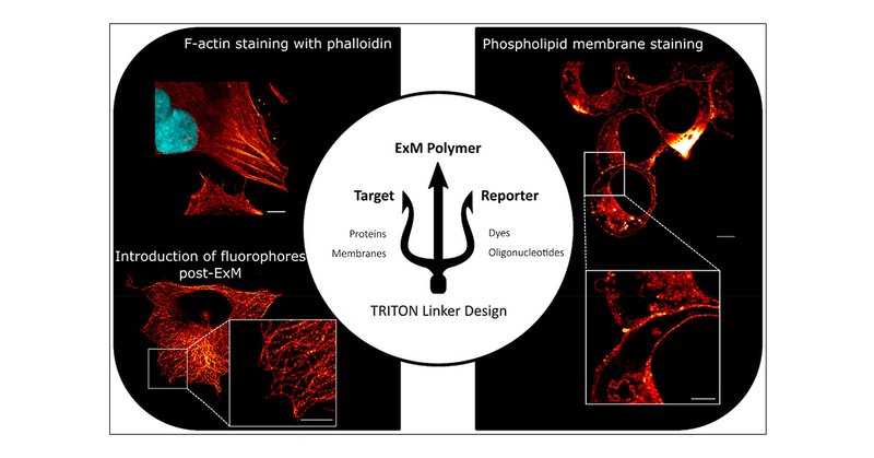
pubs.acs.org
Super-resolution fluorescence microscopy is a key tool in the elucidation of biological fine structures, providing insights into the distribution and interactions of biomolecular complexes down to...
1
1
5