
Jonathan Liu
@jonliu123
Followers
1K
Following
2K
Media
21
Statuses
267
@UW Professor, Director of Molecular #Biophotonics Lab. Open-top #lightsheet, #invivomicroscopy, #3Dpathology, Co-founder https://t.co/n1iZm1UuDu
Seattle, WA
Joined November 2013
Excited to share our vision for nondestructive 3D pathology for clinical decision support! We discuss optical, computational and translational challenges. https://t.co/qVCHAXjQ39
@adam_k_glaser @hopelessk @RederredeR @UWMadisonLOCI @anantm
4
41
176
⚡🎉 We are thrilled to introduce VORTEX, an AI-powered computational framework for predicting 3D Spatial Transcriptomics (ST) using 3D tissue images and minimal 2D ST! 🧬 By combining cutting-edge 3D non-destructive tissue imaging with AI, VORTEX imputes the 3D molecular
3
36
179

New review out on imaging 3D cell cultures. Congratulations to Huai-Ching and @jonliu123 on leading this excellent new review!

nature.com
Nature Methods - This Review discusses current 2D and 3D microscopy methods for imaging three-dimensional cell cultures and emerging strategies to address key challenges.
0
6
39
Excited to work on this with @alpenglowbio @AI4Pathology @EbenRosenthal @TopfHNS and our talented engineers and clinicians @UW !

A new project led by ME Professor @jonliu123 aims to help surgeons to remove tumors more completely & rapidly during a single procedure. The multi-university and industry partnership is funded by an up to $21.1 million award from @ARPA_H. https://t.co/Zy7uxiTCqb
4
5
27
ME Professor Jonathan Liu recently received an R01 grant from @NIDDKgov to develop computational 3D pathology methods for Barrett’s esophagus risk stratification. Learn more: https://t.co/a6M9v7C7i9
@NIH @jonliu123 @UWMedicine @BrighamWomens @fredhutch @uwengineering
0
1
10

3D tissues can reveal more about disease than traditional 2D slices, but they are enormously complex. Find out how a new AI tool can analyze these data-rich specimens to predict outcomes: https://t.co/02SE4akF0V
@GreatAndrew90 @jonliu123 @AI4Pathology @ME_at_UW
0
8
18
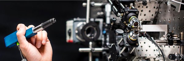
Proud of this latest publication from graduating PhD student, @Kevin_W_Bishop. In this 5th-generation of OTLS microscopes from our lab, we improve axial resolution to reduce out-of-focus background in densely labeled specimens (e.g. our fluorescent anolog of H&E).

Happy to share our latest OTLS microscope for 3D pathology from @jonliu123's lab, out now in Optics Letters! Full text: https://t.co/DOtIqC46K5
1
5
22
In addition to fully computational analyses of our #3Dpathology datasets, we are exploring AI-triage methods to keep pathologists in the loop. This should enable improved diagnostic accuracy (due to comprehensive sampling of whole biopsies) while reducing pathologist workloads.

Our paper is out @CVPR CVMI workshop! https://t.co/FwdPAnQvPh We report a DL approach that leverages contextual information along the depth axis to identify the highest-risk 2D sections within whole biopsies to help pathologists diagnose diseases more accurately.
0
0
10

In this @CellCellPress article, @AI4Pathology Faisal Mahmood, @GreatAndrew90 & @jonliu123 develop TriPath, a method for analyzing 3D pathology samples using weakly supervised AI #ASCO24
1
7
14

With #AI, it's time for pathology to go #3D
https://t.co/DxOPhiA8eJ
@CellCellPress @AI4Pathology @GreatAndrew90 @jonliu123
5
76
207
Exciting collaboration (and more to come) with @AI4Pathology and @GreatAndrew90 , just published in Cell !

⚡️📣👇Tremendously excited to share our new @CellCellPress article, where we develop TriPath, a method for analyzing 3D pathology samples using weakly supervised AI. Article: https://t.co/L2YcumCxue. TriPath enables 3D computational pathology via 3D multiple instance learning
1
0
17

#FeaturedProtocol: A workflow for robust #3Dpathology datasets of whole preclinical & clinical tissues from @Kevin_W_Bishop & @jonliu123
0
5
11

HUGE NEWS! In our new preprint, we introduce “grayscale image z-stack-guided multiphoton optical-lithography” (GIZMO) to rapidly photomodulate materials in full 3D non-binary patterns at sub-µm resolutions spanning large volumes (>mm3). https://t.co/5QJmZDschS Paper 🧵 (1/19)

Grayscale 4D Biomaterial Customization at High Resolution and Scale https://t.co/SnamjMqcUn
#bioRxiv
13
35
189
Our end-to-end workflow for 3D pathology is now published in @NatureProtocols! This includes all the steps to go from archived pathology tissues to 3D H&E-like datasets, with an emphasis on quality control for large studies. Full text at: https://t.co/MfPzUCe3aJ
3
71
267
Entertaining article by David Sokolov at Fred Hutch Cancer Center on our AI-triaged 3D pathology work led by @LindseyBarner and Gan Gao with @DeeptiReddiMD and Bill Grady… https://t.co/gMVQU7xe0W via @fredhutch

fredhutch.org
A collaboration between the Liu Lab at UW and the Grady Lab at Fred Hutch results in an AI algorithm to assist pathologists in using 3D histological specimens to diagnose esophageal cancer.
0
1
6
Super excited to see our review paper on AI for computational pathology finally out!! We provide an extensive coverage of how AI has and will shape the field of pathology. Such a fun experience with my co-author @GuillaumeJaume, and @AI4Pathology Link:
4
16
77
Check out @LindseyBarner 's final PhD paper from my lab! "AI-triaged 3D pathology to improve detection of esophageal neoplasia while reducing pathologist workloads" With @DeeptiReddiMD and Bill Grady at @fredhutch and @AI4Pathology at @BrighamWomens. https://t.co/1fzBcOzbz6
0
12
65
It’s one thing to publish images that represent our best “outliers”(which we all do initially), but a different challenge to image hundreds of clinical samples with near-100% yield. Here we release our tips and tricks from sample prep to QC, from years of iteration! #3Dpathology
Excited to share our end-to-end workflow for 3D pathology - protocol preprint is out now! More👇👇 https://t.co/82ht7ihnG1
1
7
38
Great collaboration with @GreatAndrew90 and @AI4Pathology at @harvardmed … weakly supervised learning with #3Dpathology datasets shows that block-based analysis has advantages over 2D analysis, and that performance scales with the amount of tissue volume analyzed.

Excited to share MAMBA, a deep learning computational platform for 3D pathology analysis, validated on microcomputed tomography and open-top light-sheet microscopy 3D datasets! #3dpathology #computationalpathology Pre-print:
0
1
5
Happy to report that the 3D pathology datasets from our prostate gland analysis study [W. Xie, et al. Cancer Research, 2022] are now available to all through The Cancer Imaging Archive (TCIA). #lightsheet #deeplearning #3Dpath

cancerimagingarchive.net
0
1
7