
Shalin Mehta
@mattersOfLight
Followers
1K
Following
3K
Media
142
Statuses
784
intrigued by light, logic and life. Platform Leader, Computational Microscopy at CZ Biohub (@czbiohub). Views my own.
San Francisco, CA
Joined May 2014
How do organelles change over the course of a stem cell’s life? Allen Distinguished Investigators @cohenlaboratory, @AssafZaritsky, and @mattersOfLight are using AI and cell engineering to explore the #FrontierScience of stem cell differentiation. 🔗 https://t.co/pq6AeT7loq
0
8
16
We have a fun update on DynaCLR ( https://t.co/xcCmu0bURH), a self-supervised method for learning cell state dynamics from 3D multi-channel movies! Congratulations, team @czbiohub, for reaching this milestone! Special thanks to Carolina Arias and her team for the close

arxiv.org
We report DynaCLR, a self-supervised method for embedding cell and organelle Dynamics via Contrastive Learning of Representations of time-lapse images. DynaCLR integrates single-cell tracking and...
Excited to share an update on #DynaCLR—a self-supervised method to learn dynamic cell & organelle embeddings from time-lapse microscopy using contrastive learning. DynaCLR combines single-cell tracking & time-aware sampling for robust representations. https://t.co/7llrYuZBfz
0
6
22

Virtual staining offers a promising approach to map dynamic cellular systems across various cell types + conditions, broadening the scope of live-cell imaging. Congrats to Shalin Mehta & the team at #CZBiohubSF 🎉🙌 https://t.co/kfxdF31TDV
1
39
190

🚫 No dyes. No bleaching. 🔬 Just AI + label-free microscopy = vivid virtually stained images New in @NatMachIntell: A deep learning model that enables robust virtual staining across microscopes, cell types & conditions. #CZBiohubSF
@mattersOfLight explains:
4
85
364
I am excited to join the next cohort of Allen Distinguished Investigators and looking forward to parsing the role of organelle interactions with imaging and AI in partnership with @cohenlaboratory and @AssafZaritsky!

How do organelles change over the course of a stem cell’s life? @cohenlaboratory, @AssafZaritsky, and @mattersOfLight are using machine learning and cell engineering to study the impact of organelle-organelle interactions on stem cell differentiation. #FrontierScience
5
5
54
How do organelles change over the course of a stem cell’s life? @cohenlaboratory, @AssafZaritsky, and @mattersOfLight are using machine learning and cell engineering to study the impact of organelle-organelle interactions on stem cell differentiation. #FrontierScience
2
8
39
Writer @Fairy__Hedgehog covers how microscopy researchers use AI, including @mattersOfLight, who has pioneered AI-augmented label-free light microscopy for biology at #CZBiohubSF, and #CZBiohubSF Investigator @optrickster, who uses AI for lensless imaging.

Can researchers use AI to take microscopy to the next level? From virtual staining to lensless microscopes, researchers are capitalizing on artificial intelligence to rethink microscopy. https://t.co/Vyw2ZOh1KI .
1
2
10
Thanks to students and faculty for their company and contributions. Hope to see many of you again soon!
0
0
6
Take a look at course repository for tutorials on the state-of-the-art deep learning methods for bioimage analysis:
github.com
Contribute to ai-mbl/DL-MBL-2024 development by creating an account on GitHub.
1
1
4
Directing this year's DL@MBL course with @janfunkey, @sagzehn, @ilastik_team, and our awesome TAs was one of my most intense and fulfilling projects of 2024. The spirits of curiosity, collaboration, and Captain Kidd 🙃 made the course so much fun! https://t.co/X5yb1nciC7
1
5
31
The combination of virtual staining and ultrack enables label-free segmentation and tracking: https://t.co/RPoupikQSl This has already enabled gentler imaging and analysis of cell dynamics in our projects.

7/ 🤖 As mentioned, we can also directly use the intensity images if they already delineate the cells with contours, as is often the case with membrane labeling. With the help of @mattersOfLight 's team, particularly @edyoshikun, we tracked A549 cells using quantitative phase
0
0
1
Shout out to @jobragantini and @loicaroyer for leading this effort to develop a versatile cell tracking method. Check it out!
📡🧵 Excited to release our preprint: Ultrack: Pushing the Limits of Cell Tracking Across Biological Scales https://t.co/R7iXuhEGUH A tour-de-force by @jobragantini, #Ultrack is a versatile, highly accurate, and fast ILP-based cell tracking software for 2D, 3D, and multicolor
1
0
6
1/ 📡Exciting news! 🚀 Our #Omega paper has been accepted at @NatureMethods! Omega is a @napari_imaging plugin that uses Large Language Models to assist users with bioimage processing and analysis tasks! Check out our original project announcement (see below), read the paper:
🚨 #ChatGPT + @napari_imaging 🚨 Releasing my latest weekend project: Omega, an autonomous LLM agent that writes image processing and analysis code, fixes its mistakes, accesses the napari viewer, makes widgets, & more! https://t.co/5btZq05ro1
@LangChainAI @OpenAI
#OmegaAgent
8
60
256

I've been so out of breath trying to deal with grant deadlines that I've barely had time to pause and appreciate the massive amount of progress we made in the last year in spite of the move from Salk to UCSD and hanging around in temporary space (permanent lab is done 7/31!) 🧵/1
5
4
80
Finally, thanks to Priscilla Chan and Mark Zuckerberg for their support, and to the leadership at @czbiohub for creating an environment where partnerships between technologists and biologists are the norm. /end
0
0
7
Successful execution required the integration of ideas across optics, algorithms, automation, high-performance computing, cell biology, and neuropathology. I am thankful to my colleagues for their expertise and for placing this bet with me. 14/n
1
0
6
@czbiohub @LiHao_Yeh @orctweets This is one of those projects that has taken several iterations and years to finish - look at the timestamps! Special thanks to @rita_strack for being an amazing editor throughout this process. 13/n
1
2
10
@czbiohub @LiHao_Yeh We needed a calibration target to evaluate the measurements of 3D dry mass and 3D orientation made with PTI. In addition to biological structures of known architecture and isotropic beads, we used anisotropic glass targets built by the Peter Kazansky lab @orctweets. Intriguing
1
0
3
@czbiohub As I mentioned before, the design complexity lies in the algorithms and calibration. @LiHao_Yeh validated the wave optical model of the microscope and the reconstruction algorithm with methodical simulations of isotropic beads and an anisotropic patterns. 🌊💡 11/n
1
0
2
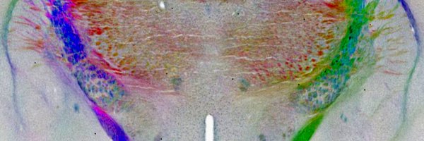
@czbiohub PTI is easy to multiplex with fluorescence and H&E imaging, allowing label-agnostic mapping of cell and tissue architecture. In this movie, we image a standard H&E stained cardiac tissue slice. 10/n
1
0
2