Pulmonary-POCUS
@HoosierPocus
Followers
2,441
Following
237
Media
178
Statuses
636
Pulmonary and Critical Care Medicine Point Of Care Ultrasound (POCUS) education. By @Edwin_J_Jr Website coming soon
Indianapolis, IN
Joined May 2022
Don't wanna be here?
Send us removal request.
Explore trending content on Musk Viewer
#KAZZAWARDS2024
• 1063811 Tweets
SAROCHA REBECCA IN KAZZ
• 767058 Tweets
FOURTH x KAZZ 2024🥳
• 128118 Tweets
#Aぇǃgroup
• 106551 Tweets
ナイジェリア
• 58104 Tweets
#お迎え渋谷くん
• 49871 Tweets
インプレゾンビ
• 48326 Tweets
Varane
• 47452 Tweets
로즈데이
• 33994 Tweets
groupデビュー
• 30724 Tweets
#MEOW_HAERIN_DAY
• 27135 Tweets
#芳典に夢中な私はもうSTUPID
• 26947 Tweets
MAY THE 24TH BE WITH YOSHI
• 26390 Tweets
HAERIN SWEET 18
• 20117 Tweets
Powell
• 16793 Tweets
CLIPE DE ACEITA
• 16374 Tweets
ジュニア最後
• 12017 Tweets
Nicolas Cage
• 11941 Tweets
BOARD THE WISHBUS WithJC
• 10307 Tweets
ホットケーキ
• 10217 Tweets


Pinned Tweet

4
#POCUS
Questions of the week?
1. What view is this?
2. How is the view made?
#howdoimakethatview
3. Can you name the structures?
4. What can be recorded from this view?
#IUPCCM
,
#PulmonaryPOCUS
#howdoImakethatview
,
#ECHOfirst
,
#RDCS
,,
#FOAMed
,
#IMPOCUS
7
31
101
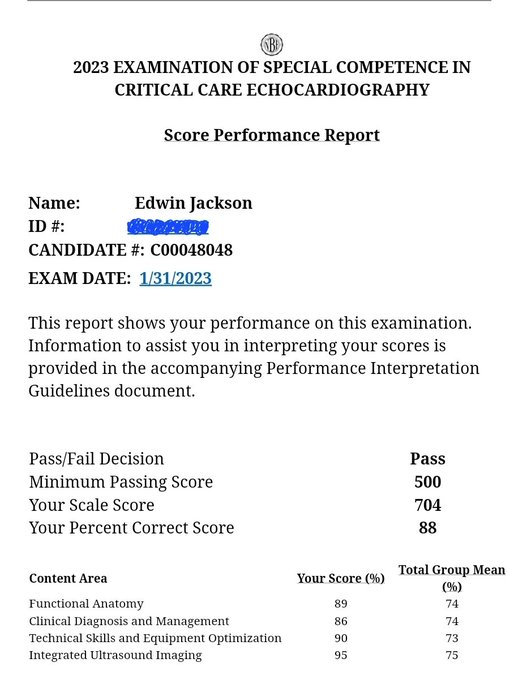
#POCUS
question of the day?
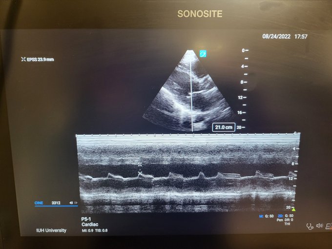
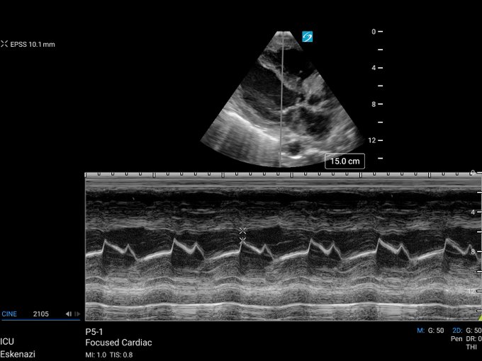
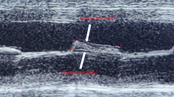
1. What does this image show, and what mode of ultrasound was used to obtain it?
2. What is the E-point?
3. What is the A- point?
4. What is EPSS, and why do we use it?
Scroll down to learn how to interpret the image.
5
72
208
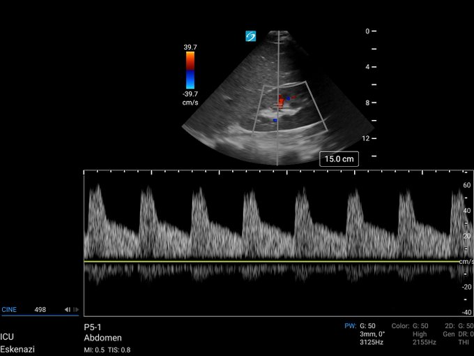
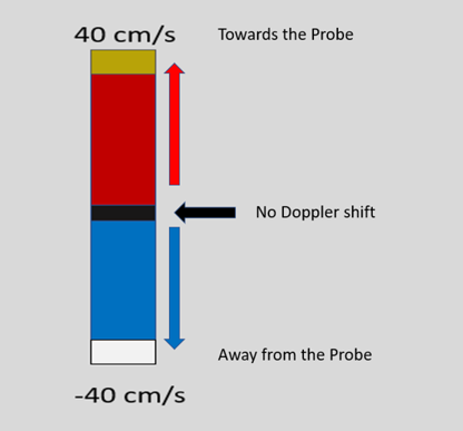
#POCUS
, Color Doppler
1/6
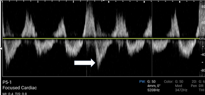
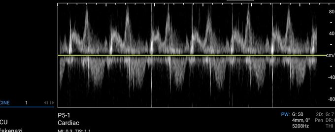
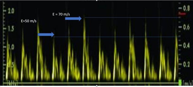
Q1: What color is assigned to blood flowing toward the probe at 31 cm/s in this image?
Q2: What color is assigned to blood flowing away from the probe at 18 cm/s in this image?
Edelman Understanding US Physics 4th Edition.
3
25
115
#POCUS
More on EPSS
#howdoimakethatview
1. Obtain a parasternal long-axis view
2. Select M Mode
3. Place the M Mode line through the mitral valve leaflet tips
4. Select M Mode again.
5. Select Freeze
6. Select Calculations, Packages, Cardiac, Function, EPSS
1
26
74
1/2
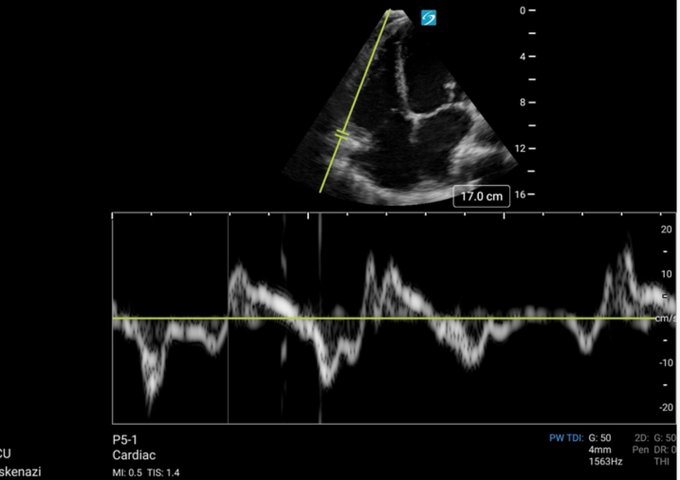
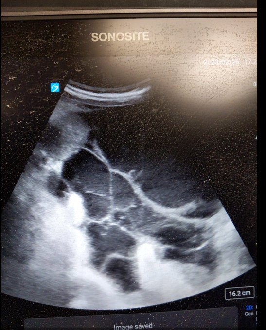
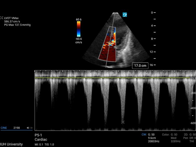
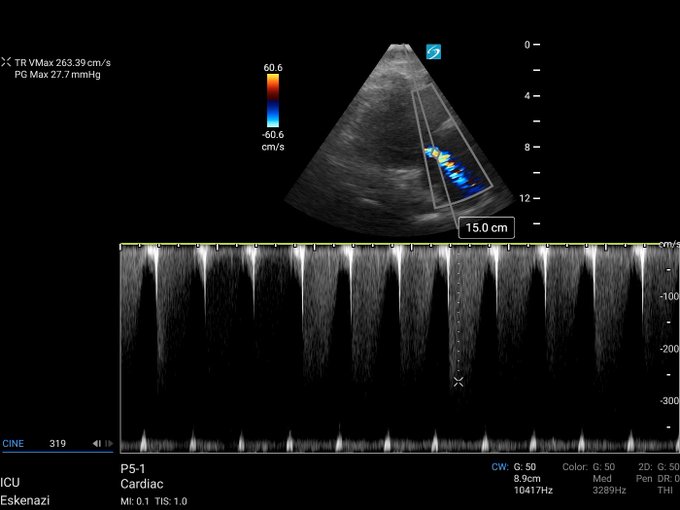
#POCUS
Critical Care Echo
The following images were obtained from a 50-year-old male with refractory hypotension in the ICU. PW Doppler was placed through the LVOT with significant aliasing. See the CW Doppler on 2/2
Name 3 structures?
What's your differential diagnosis?
6
26
73
Name two valvulopathies in this clip.
What view is this?
What adjustments can be made to improve this image?
#POCUS
@NephroP
@GrahamCarlos
@IUPCCM
@zedunow
11
21
68
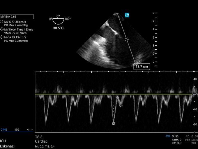
When planning a thora or percutaneous chest tube how many of you use color Doppler to look for vessels?
9
14
65
5
8
46
#POCUS
Extra, extra credit
Our last tweet showed dynamic LVOTO; here is a clip of the mitral valve Color Doppler in the same patient.
Q1: What finding is shown here?
Q2: Is his finding related to dynamic LVOTO?
@NephroP
@TaweevatA
@OSUPCCM_Fellows
@IUPCCM
@IUEM_ultrasound
7
15
43
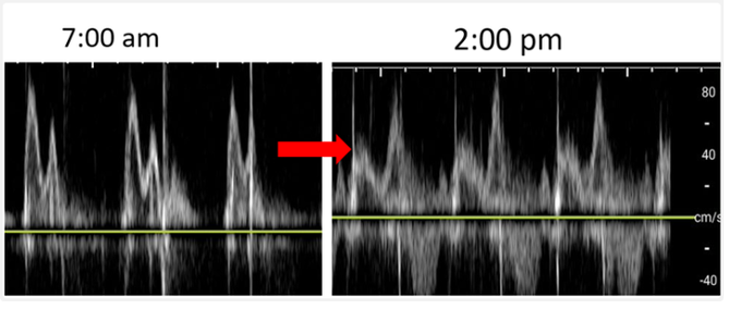
A patient is admitted for dyspnea; you note that he had a TTE 3 months ago with normal LVEF, LA dilation, and stage I diastolic dysfunction. He was given 2 L of fluid and was subsequently intubated. What intervention was performed between 7:00 am and 2:00 pm? See next post
6
7
40
1/2
A 45-year-old female with a history of polysubstance abuse presents with a cough, purulent sputum, & fevers. Blood cultures are positive for GPCs. CXR shows a dense left lower lobe infiltrate. Describe the findings below.
@NephroP
@IUPCCM
@OSUPCCM_Fellows
2
9
39
R IJ CVC placed followed by 10 ml of saline injected through the distal port.
What sign is this?
Why are the ventricles upside down?
@ProcedurePRIME
@NephroP
@BModeBowling
6
6
39
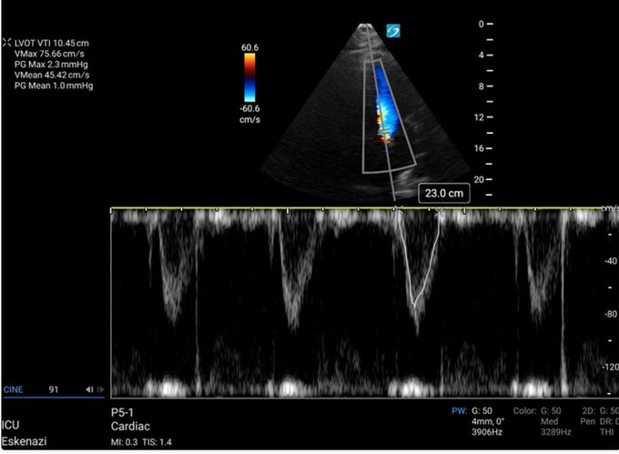
POCUS Clip and Quiz of the Week # 28
The LVOT diameter is 2.0 cm, and the LVOT VTI is below (10.45 cm). The patient's HR is 80 beats per minute; what is the cardiac output?
Find out by taking this week's quiz--->
@IUPCCM
@GrahamCarlos
@NephroP
2
8
37
Attention ultrasound experts! Please post your most impressive aortic valve image. Let's highlight POCUS in action!
#AorticValveViews
#POCUS
@NephroP
@ross_prager
@argulian
@BModeBowling
7
9
38
What valvular abnormality is being shown here?
What can be done to improve your ability to assess valvulopathies?
Want to find out? Join the
@IUPCCM
by taking this week's clip/quiz.
@NephroP
@GrahamCarlos
@ForeverFancy17
@OSUPCCM_Fellows
6
6
37
Quiz of the Week 27
You are called to evaluate a 47-year-old male with a history of antiphospholipid antibody syndrome. He has tachycardia and chest pain. What is your diagnosis? Find out by joining the
@IUPCCM
in this week's quiz
@GrahamCarlos
@NephroP
6
9
37
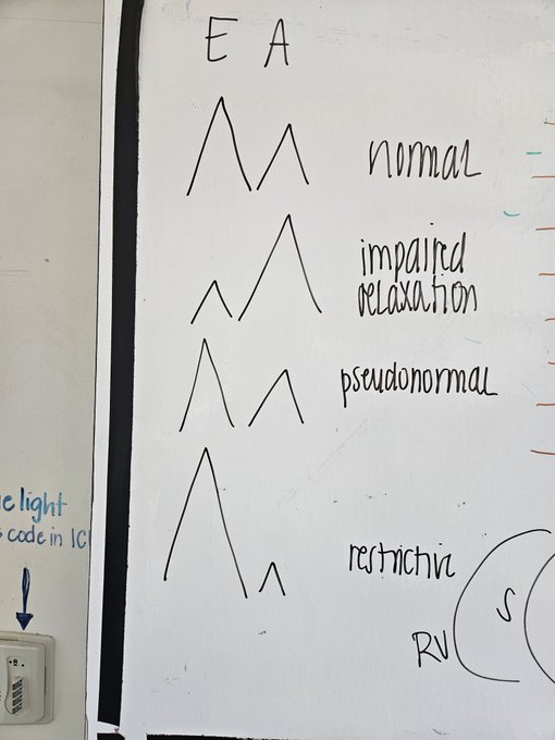
I walk into the team room on Friday and find the chalk talk of our fantastic fellow
@juliehcarroll
and our awesome residents. The mitral inflow pattern and its association with left atrial pressure is key in the ICU
#POCUS
.
@IUGenMed
@IUPCCM
@NephroP
@GrahamCarlos
@tjelle13
1
8
36
Join the IUSM PCCM Fellows and take the POCUS Quiz of the week. Click the link below!
0
8
36
It's time for Quiz/Clip of the week # 15
What view is this?
What LV walls are seen here?
Find out by joining the
@IUPCCM
and take this week's quiz.
Xtra credit: Comment on the RV size
-->Link
@NephroP
@ForeverFancy17
@GrahamCarlos
@OSUPCCM_Fellows
9
13
35
What type of Doppler is this?
What is being measured here?
Would you consider this normal or abnormal?
Find out by joining the
@IUPCCM
and take Clip/Quiz of the week
#20
@ForeverFancy17
@NephroP
@GrahamCarlos
#POCUS
#TDI
12
8
34
What measurement can be made here?
Find out by joining the
@IUPCCM
and taking quiz # 18
Link-->
@GrahamCarlos
@NephroP
@ForeverFancy17
@ross_prager
11
6
34
2
4
34
72 y/o female presents with dyspnea; she has a history of renal cell carcinoma. US of the left lung shows
@IUPCCM
@OSUultrasound
@OSUPCCM_Fellows
@IUEM_ultrasound
@IUIntMed
@NephroP
5
12
34
Amazing view, should be a mainstay in the ICU. I find pulmonary artery acceleration time much easier to get in this view.

In patients with difficult parasternal windows, especially COPD, subcostal short axis is commonly a rescue. In this patient, note partial fusion of RCC and LCC, as well as good delineation of the pulmonic valve
10
56
285
0
3
32
1/7
#POCUS
Questions of the week
1 What is RVSP & what valvular velocity is required for its calculation?
2 What views are used to calculate RVSP?
3 How is the view made?
#howdoimakethatview
4 What two types of Doppler are required?
5 What equation is used to calculate RVSP?
2
12
33
In the ICU today, pt with acute shock and hypoxia
What do you see here?
What's your diagnosis?
@IUMedSchool
@IUGenMed
@IUPCCM
@NephroP
@GrahamCarlos
@ForeverFancy17
8
17
33
"Take a look at this image from a transcranial Doppler. Can you identify the vessel being shown? 🧠🔍
#BrainVoyage
#POCUS
TCD
@NephroP
@IUPCCM
@ross_prager
3
5
30
Image of the day, this is a ultrasound of the left upper quadrant in a patient in profound septic shock. What's your differential?
6
4
30
What view is this?
What do you see in this clip with color Doppler?
Can you make this view?
#POCUS
@GrahamCarlos
@ForeverFancy17
@IUPCCM
@NephroP
@OSUPCCM_Fellows
3
9
29
2/2
Additional information: The patient was on 30mcg/min of NE, IVC 1.1 cm with >50% respiratory variation (not intubated).
Extra credit: how should this be managed?
4
9
29
Which leaflets of the tricuspid valve are seen here?
Find out by joining the
@IUPCCM
by taking this week's quiz.
#POCUS
,
#echofirst
@GrahamCarlos
@NephroP
@POCUSpeek
@IUIntMed
@IUEM_ultrasound
@IUGenMed
@OSUPCCM_Fellows
4
8
26
What view is this?
What's in the RA?
@IUPCCM
@OSUPCCM_Fellows
@CCF_PCCM
@GrahamCarlos
@NephroP
@ross_prager
@khaycock2
@Srivatsa34
2
6
27
What's your diagnosis?
Which is the true lumen?
@NephroP
@GrahamCarlos
@ForeverFancy17
@Srivatsa34
@TaweevatA
6
7
27
After a long 12 weeks of waiting the results of the CCEeXAM are out. This was a very challenging examination and I learned a lot of echocardiography studying for it. Thank you too
@IUPCCM
for giving me the time and resources and thank you too
@OSUPCCM_Fellows
as well!
8
1
27
85-year-old male with a known malignancy presents with this finding.
1. What is the most common malignancy that presents this way?
See second tweet for additional information
7
10
26
What view is this? Can you comment on this patient's diastolic function with the knowledge that their e' velocity is 4 cm/s and their TR max is 320 cm/s?
Is the LAP high or low?
@ross_prager
@NephroP
@POCUSpeek
@IUPCCM
@OSUPCCM_Fellows
@POCUSpeek
@RaniaRightHeart
2
2
25
What view is this? I really like using it in my COPD patient when I want to interrogate the pulmonary artery.
@NephroP
@Srivatsa34
5
8
25
Great image from
@NephroP
. Looking at this left ventricle what is the major risk factor for the development of the thrombus?

4
61
269
0
6
25
What organ is being shown in this clip and what vessesl are being interogated? Want to find out? Join the
@IUPCCM
and take this weeks quiz.
@NephroP
@ATSMedEd
@GrahamCarlos
2
4
24
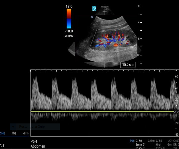
Quiz/Clip of the Week # 23
This spontaneously breathing patient has a right atrial pressure of 10 mmHg. What is the RVSP if the TR max is 344.08 cm/s?
What equation should be used here?
Join the
@IUPCCM
and take this week's quiz.
@NephroP
@Srivatsa34
3
9
25
@juliehcarroll
performed her first cTEE today. She did an excellent job, and made an unexpected diagnosis!
@IUPCCM
has some amazing fellows!
@gbosslet
@RFPMachado
@GrahamCarlos
@LauraHinkle18
;Joe Smith for the support!
@NephroP
@ross_prager
, can anyone name the view?
2
3
22
"Curious about your POCUS prowess? 🤔 Can you explain how the transducer was manipulated from the first image to the last? Share your insights! 🩺🔊👀
#POCUS
#ImageAcquisition
"
@NephroP
@BModeBowling
@Srivatsa34
@Irina67790690
0
4
23
Does the type of Doppler in this clip have range specificity?
Is blood flow moving towards or away from the transducer?
Want to find out? Join the
@IUPCCM
and take this week's quiz.--->
@ForeverFancy17
@GrahamCarlos
@POCUSpeek
1
5
22
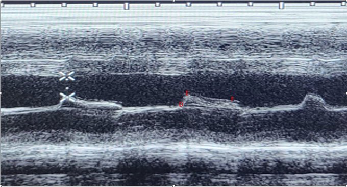
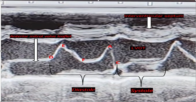
Extra Credit
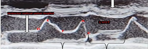
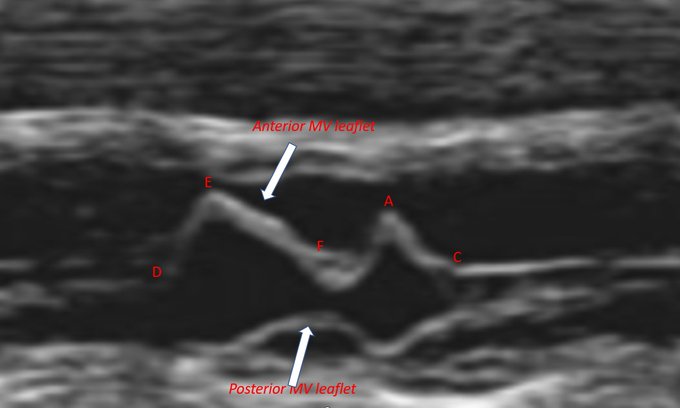
F Point: marks the onset of diastasis slow flow
C Point: The point where the anterior and posterior leaflets of the mitral valve come together during systole.
D Point: Mitral valve opening at the beginning of diastole
1
4
23
We're zooming in on a unique view – but what exactly are we seeing? 🤔 Notice the echogenic structure inside the vessel at the 9 o'clock position. 🕘 Can you guess what it is? What 3 vessels are we seeing?
@NephroP
@ross_prager
@IUPCCM
4
3
21
@NephroP
Look at that descending aorta it's huge and there's a dissection flap inside of it. Pretty rare to catch it like that in my opinion.
Great image!
0
1
21
How many structures can you name? Join the
@IUPCCM
and find out by taking this week's quiz! Just click the link below!
#CCE
,
#POCUS
@OSUPCCM_Fellows
@IUSMDeptMed
@GrahamCarlos
1
7
21
The hits keep coming! Another awesome post in example of dynamic LVOTO
@IUPCCM
@ForeverFancy17
@GrahamCarlos

(SAM) is displacement of the distal of anterior leaflet of mitral valve toward the LVOT. SAM can occur in patients without (HOCM) and is a well-recognized cause for unexplained hypotension in perioperative settings
#echofirst
#echo
#CardioTwitter
#cardiovascular
#HCM
#Cardiology
0
22
50
1
4
21
Clip of the week
#30
This week, we will explore the abnormal hepatic vein Doppler flows frequently encountered in critical care settings.
What wave is the white arrow pointing to? Find out by taking this week's quiz ---->
@IUPCCM
@GrahamCarlos
1
3
20
5/5
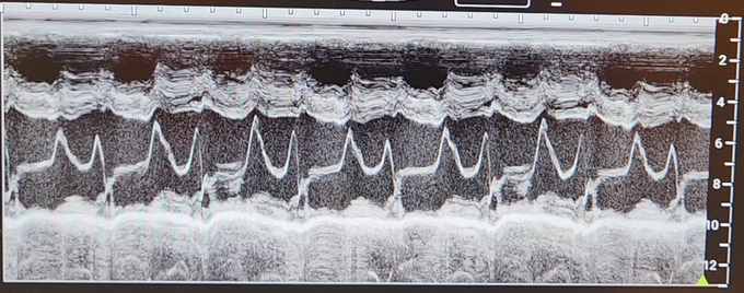
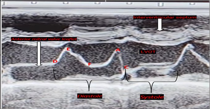
The M-mode combination of leaflet thickening, the anterior motion of the posterior leaflet, a flat E-F slope, the absence of a distinct A point, and decreased D-E excursion are very suggestive of mitral stenosis.
Extra credit: Can MV stenosis severity be determined by Mmode?
2
3
20
3
6
19
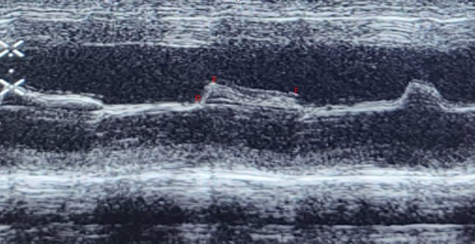
Answer 1: The image above shows the mitral valve in M mode taken during the PLAX view at the MV leaflet tips.
Answer 2: During early diastole, the leaflets separate widely with the maximum early diastolic motion of the anterior leaflet termed the E-point

1
3
20
3/5
Remember what normal looks like?
D = Mitral valve opening
E = Maximal opening during early rapid filling
F = Onset of diastasis
A = Atrial contraction
C= Mitral valve closure
1
5
19
7
8
19
@fersaurin
@NephroP
@IUEM_ultrasound
@IUPCCM
@IUIntMed
@OSUPCCM_Fellows
Glad you asked! This paper by Feigenbaum is by far the best summary of M mode IMO.
Role of M-mode Technique in Today's Echocardiography
2
5
19
This is a very important concept to understand!
@NephroP
thanks for sharing!
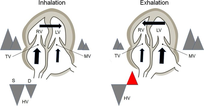
Respiratory variations in constrictive pericarditis.
Click 'ALT' for figure description. TV = tricuspid valve Doppler MV = mitral valve HV = hepatic vein
#VExUS
#POCUS
#echofirst
#MedEd
🔗
0
56
136
0
3
19

POCUS Quiz of the week!!!
What is this view?
Can you name the papillary muscles in this view?
Join the
@IUPCCM
fellows and take this week's quiz by clicking the link below!
@NephroP
@POCUSJournal
@POCUSpeek
@GrahamCarlos
@ForeverFancy17
1
4
18
Picture perfect PISA
@VLSorrellImages

This may be the BEST reverse PISA (for mitral stenosis) ever seen.
Thanks to
@swleun2
for these phenomenal images!
#EchoFirst
#JADEL
@ASE360
14
64
260
0
5
17
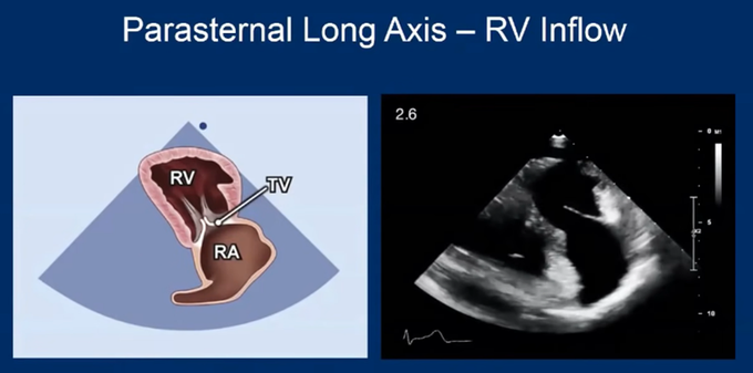
Answer 3. The RA cavity, tricuspid valve (anterior and posterior leaflets), coronary sinus entry into the RA, and the RV inflow up to the apex of the RV can be seen in this view.
1
3
18
What do you think about this patient's left atrial pressure,--> low/normal or high? And why?
@TaweevatA
@NephroP
3
1
18
It's that time again! Quiz/Clip of the Week # 16
What view is this?
Which LV walls are seen in this view?
Can LVOT-VTI be measured here?
Join the
@IUPCCM
and find out by taking this week's quiz-->
@NephroP
@ForeverFancy17
@POCUSpeek
3
8
18
Answer 4: EPSS = E-point septal separation and is defined as the distance between the anterior leaflet of the mitral valve and the interventricular septum.
1
3
17
Answer 4. This view is suitable for recording the velocity of tricuspid valve regurgitation. The jet is often not parallel to the color flow, which can result in underestimating the TR max. TR should be evaluated from A4C, PSAX & RV inflow.
1
2
18
2/5
What is happening to the E-F slope?
What is happening to the D-E excursion?
1
1
17
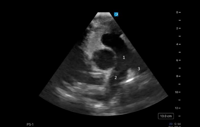
How many structures can you name?
Join the
@IUPCCM
by taking this week's quiz.
QR code in the second tweet.
@NephroP
@OSUPCCM_Fellows
@IUMedSchool
@IUIntMed
#POCUS
2
1
16
@NephroP
The arterial line indicates increase ventricular interdependence. With inspiration the RV preload is increased thus pushing the interventricular septum into the LV decreasing volume and dropping systemic blood pressure. This patient could have tamponade or COPD etc..
1
1
16
@juliehcarroll
and I needed to have a look at these structures in our post-cardiac arrest patient, can you name them? Can you name this view?
#ccTEE
#echofirst
@NephroP
@ross_prager
@ThinkingCC
@IUPCCM
3
7
16
1/2
Can you name this view?
How is this view made?
Find out by joining the
@IUPCCM
by taking this week's quiz.
Click here-->
@NephroP
@GrahamCarlos
@OSUPCCM_Fellows
@POCUSpeek
4
6
15
@NephroP
Can't use IVC diameter or variation with a sniff on positive pressure. I treat the NIPPV the same as an ET tube in this instance. If IVC diameter is less than 1.2 cm, RAP is less than 10. Could use s',CVP or the SVC if you can image that w/o TEE. Interested to hear from others
2
1
15
Answer: Septated (L) pleural effusion imaged posteriorly with a phased array transducer at the left scapular line. This view provides a short axis cardiac window. Thora revealed a complicated parapneumonic effusion.
@GrahamCarlos
@ATSMedEd
@NephroP
@ThinkingCC
@IUMedSchool
0
5
16
What view is being made?
What is in the left atrium?
Want to find out?
join the
@IUPCCM
fellows and take this week's quiz
@NephroP
@GrahamCarlos
@ForeverFancy17
@Srivatsa34
#POCUS
1
2
15
Next week we will review how we can use TR Max to calculate the right ventricular systolic pressure.
Extra Credit: The RV inflow view can also confirm central lines by using the rapid swirl sign.
#CCPOCUS
,
#PulmonaryPOCUS
#RDCS
,
#FOAMed
1
2
16
4
6
14
Question of the day
#1
:What pathology is seen here?
#2
: Where is the transducer located?
#3
: What procedure should be performed?
@NephroP
@GrahamCarlos
@ATSMedEd
@Srivatsa34
@IUPCCM
@IUMedSchool
#Echofirst
#CCEcho
#pleuraleffusion
4
4
16
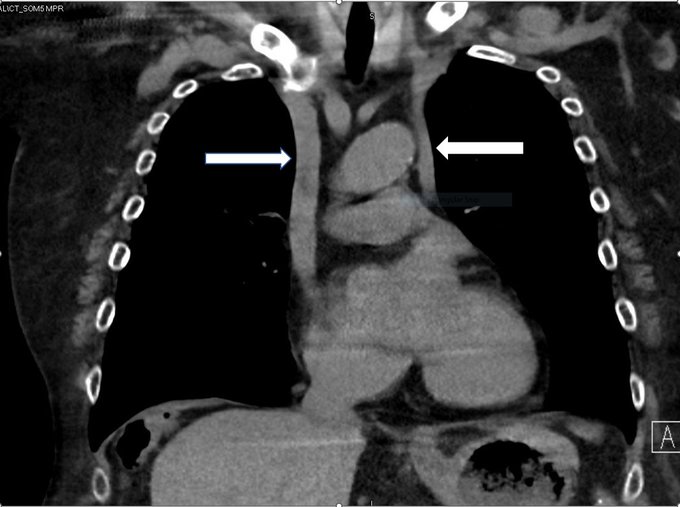
You see this coronal CT image of a 50 y/o female as you are preparing to place a L IJ CVC.
What can you tell me about the probable size of her coronary sinus?
What is the diagnosis?
How is the diagnosis made?
@GrahamCarlos
@NephroP
@IUPCCM
@ForeverFancy17
@OSUPCCM_Fellows
4
2
13
A young man presents to the ED with 2 weeks of left-sided chest pain, fevers, and cough with purulent sputum. He was treated for CAP 2 weeks ago for an apparent LLL pneumonia. POCUS exam of the left chest shows
What is that?
#pleura
3
6
14
.
With a normal healthy heart, the tip of the anterior mitral valve leaflet nearly or completely encounters the interventricular septum. In a low cardiac output state the MV does not have to open very wide to blood from the LA to LV. Notice how the MV touches the IVS.
1
6
14
What's the over under here?
@katiewiskar
can you repost that clip of the vascular bundle on top of the rib?
5
4
14
Here are a few answers to this week's quiz. Click on the link to learn more about the aortic valve and LVOT.
#POCUS
0
2
14
What's the diagnosis? Which mitral valve scallop is this, hint it's located near the left atrial appendage? 🌊🐚
#CriticalCareTEE
@ross_prager
@NephroP
@_Azaay
@IUPCCM
@emireles_c
@KalagaraHari
5
4
13
What is the white arrow pointing to?
What part of the LV is the red arrow pointing to?
Want to find out? Join the
@IUPCCM
and take this week's quiz!
Click the link below
2
6
14
1
5
13
Quiz week # 26
Does this patient with a large pericardial effusion have ventricular Interdependence based on the transmitral velocities pictured below? Is the sweep speed high or low? Find out-->
@IUPCCM
@NephroP
@Srivatsa34
1
6
13
3
6
13
2
2
13
That's an amazing image!

2 beauties in one shot! BAV + myxoma on TEE.
#echofirst
Friday!
@VLSorrellImages
@ChetritMichael
@sarah_blissett
@ASE360
@RodrigoBagur
@HeartDocSharon
5
25
139
0
0
13
4/5
Our patient's MV (Left) has poor D-F excursion and a flat E-F slope. The posterior MV leaflet is also being pulled anteriorly. An example of normal MV M-mode motion is seen on the (right)
1
2
12
What view is this?
What coronary arteries can be assessed here?
Find out by joining the
@IUPCCM
by taking this week's quiz. Click the link
@GrahamCarlos
@ForeverFancy17
@IUIntMed
@NephroP
3
3
12
A 55-year-old male presents with chest pain after a fall 24 hours ago. You perform lung US and save the following clip. What is your diagnosis?
Join the
@iupccm
fellows and take this week's quiz to find out!
4
5
12