
Johns Hopkins Pathology
@JHUPath
Followers
14K
Following
226
Media
340
Statuses
942
The Department of Pathology at Johns Hopkins University
Baltimore
Joined August 2010
Applicants must: Hold an M.D. degree Be pursuing or have completed residency or fellowship training in anatomic and/or clinical pathology Be willing to commit to a minimum of one year in the program Be eligible for NIH training grant support
1
0
0

1/ A high-functioning board is the ultimate leverage for a founder. Here’s why (and how to build one):
10
15
175
The program is suited for pathologists who aspire to become principal investigators leading laboratory-based research groups, and may be particularly attractive to those interested in conducting research at Johns Hopkins during or following their clinical residency or fellowship.
1
0
0
Selected research fellows will join laboratories at the Johns Hopkins University School of Medicine and participate in coursework and structured activities to introduce trainees to topics in cancer research and fundamentals of academic career development.
1
0
0
We are Accepting Applications for the Opportunities for Pathology Trainees in Cancer Research (OPTIC) Cancer Biology Research Training Program at @HopkinsMedicine , funded by @theNCI.
1
0
0

"If they can get him to click like they have Cade Horton, the sky's the limit." @LanceBroz does a deep dive on Cubs pitching prospect Jaxon Wiggins 👀
6
9
262
This Updates in Bone and Soft Tissue Course will be 🔥 🔥 🔥 🔥 🔥 Rome 🇮🇹 February 6-7, 2026 Come for the 🍕 🍝 🍷 or the 💀 🩻 🍖 🦴 #BSTPath
#sarcoma
#MSKRad
#orthopaedics
#pathology
#surgpath
#surgicalpathology
#bonepath
#softtissue
2
35
67

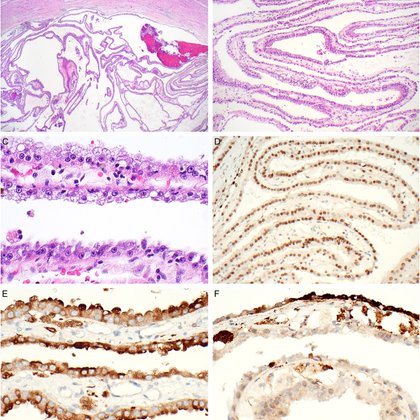
Excited to share our new @Histo_Journal correspondence on low‐grade sarcomatoid thyroid‐like follicular renal cell carcinoma - the first molecularly confirmed case showing a shared clonal origin of both components. https://t.co/8BrG47snjV Grateful to Dr. Argani @PeterArgani for
1
2
18

Study in @PNASNews ( https://t.co/1S0Yc63K3s) by @HopkinsMedicine @JHUPath shows in mice that 2 @US_FDA-OK'd pain drugs also inhibit nerve signaling/blood vessels in #Osteosarcomas & slow growth & spread. Info: https://t.co/W7VGJSbAXV.
@sowmya_rames @Qin_Qizhi @AaronWJames_Lab
2
4
7
We are inviting applications for a full-time, tenure-track SURGICAL PATHOLOGIST faculty position at the Johns Hopkins Hospital. The ideal candidate would have an area of expertise, with priority given to breast pathology @TheISBP If interested, apply at
0
3
5

gm. Love this OCM Genesis. A Cheetah fur with Bitcoin background, orange pocket square, and matching blues.
22
18
109
Johns Hopkins Pathology is very excited to host Haider Warraich for virtual grand rounds on Monday Sep 8. He'll be discussing his innovative work at ARPA-H and medical AI. Please join us online!
1
7
13
John Hopkins Pathology has an unexpected opening for our 2026-2027 advanced surgical pathology fellowship, which offers trainees the opportunity to independently sign out cases in a highly supported setting. See https://t.co/omgWnNt3t6 for more details.
0
16
22
We are excited to announce our open house Thursday, August 28th! Join us to learn more about our program, our beloved city of Baltimore, and get to know our residents and faculty! 🔬🦀 Click the link in our bio to RSVP! #pathology #residency #pathmatch25 #johnshopkins
0
16
41
The Johns Hopkins GI/Liver Pathology Fellowship has an unexpected opening for 2026-2027. Visit our website for more details and to apply: https://t.co/6vQS8cH4SB
#GIPath #pathology
0
4
5

Here comes a new challenger! Naoto Kurogane joins the dive! Specializing in close-quarters combat, Naoto’s signature ability lies in his unique power to draw upon his own blood and transform it into a deadly arsenal of weapons. Naoto can enter a special empowered state that
9
119
528
Ossifying Spindled and Epithelioid Tumor: A Novel Soft Tissue Tumor | @ModernPathology @JMcMahonG @jonathandudley @LisaRooperMD @PeterArgani
https://t.co/Fy1PHmMu83
1
12
34
Our Residency Program Ranks #1! Over 30k physicians participated in a survey to nominate residencies that offer “the best” clinical training. We are proud that once again, Johns Hopkins ranked as the #1 residency program in Anatomic & Clinical Pathology! https://t.co/LrG99pKJ6b
doximity.com
Find the best residency program for you. Read reviews and see ratings from program alumni.
0
8
39
Join Us for the 25th Annual Current Topics in Gastrointestinal and Liver Pathology CME Course! For more details and to register, please visit the link below:
0
0
2
Clinically Sporadic Folliculin-mutated Renal Epithelial Neoplasms Represent a Mixture of True Somatic Folliculin-mutated and Occult Birt-Hogg-Dubé Syndrome-associated Cases | @PeterArgani @DrSwatiBhardwaj @AJSPjournal
https://t.co/l2WdcJoTrX
journals.lww.com
radic tumors and harbored FLCN mutations and no other genetic alterations characteristic of another established subtype. On further workup, 5 seem to harbor true somatic FLCN mutations, whereas the...
0
4
16

Dr. @lauradelongwood's team identified P4HA1 as a driver of #PDAC #organoid invasion in hypoxia. @JHUPath 🕳️ Dig into the details of the study in @CRC_AACR: https://t.co/sbRwGYvWfd
0
4
18
We were so honored to welcome back @JHUpath pathology resident alumna Dr. Diana Molavi for her annual intern conference session on Bone and Soft tissue…with an impromptu book signing of “The practice of Surgical Pathology”! 😉 #pathtwitter #pathX #path2path #pathmatch25
0
3
45

Introducing BMNG, the 2x Long BMNR Daily ETF. - NO margin account needed - 0.75% Expense ratio - Nasdaq-listed Investment involves significant risk. Not a direct stock investment. See prospectus for additional details on our website.
4
14
51
MEIS1::NCOA1 Primitive Spindle Cell Sarcoma of the Kidney - Report of 7 Cases of a Distinctive Clinicopathologic Entity | @JMcMahonG @Andres_Matoso Newly published in @AJSPjournal
https://t.co/bmFXr9WIlm
1
9
27