

Alexandre Dumoulin @axonsalex.bsky.social
@AxonsAlex
Followers
1K
Following
1K
Media
69
Statuses
385
Developmental Neurobiologist @UZH_Science | Microscopy Enthusiast - find me on Bluesky: https://t.co/akB7r28gPa -
Axonal growth cone
Joined August 2020
Finally out! 🥳We added a lot of rescue experiments. We now show that Arl13b needs to locate to commissural neurons primary cilium for proper rostral turning of axons. We also further characterized Shh retrograde transport to the soma of comm. neurons.

Delighted to share our latest preprint entitled « A cell-autonomous role for #primarycilia in long-range commissural #axonguidance ». We propose a mechanism involving the neuronal primary cilium during axon guidance at an intermediate target. 🧵(1/7)

2
7
44
Here’s an embryonic axonal growth cone labeled with PK Mem dye (red) and tubulin (blue), imaged every 2 seconds. 🤩.
0
0
3
Thrilled to collaborate with @ZhixingChen2 Lab @PKU1898!.We tested new PK Mem dyes—gentle, photostable tools to label the plasma membrane in live neurons, making it easier to track growth cone motility & axonal transport. #liveimaging #neuroscience .
1
8
39
This time-lapse captures 17h of axonal growth from a chick dorsal root ganglion explant, seen through the actin cytoskeleton using live imaging. I just submitted this video to the Nikon Small World in Motion competition. Today is the last day to upload yours!😉 #neuroscience
12
124
674
RT @Ruth_Y_Williams: Come and join the Williams lab @OfficialUoM @FBMH_UoM @The_MRC We are using single-cell Multiomics and in vivo C….
0
27
0
RT @Prof_Lundberg: I’m very excited to finally present something we’ve been working on for the past couple of years – a spatial proteome ma….
0
35
0
RT @MicrobiomDigest: The punishment for science misconduct is …. nothing. All these men are still running labs / paid professors.
0
895
0
RT @frogwranglers: Baby you’re a fiiiiirework!!!. Explanted Xenopus neural crest cell, microtubules (green) and actin (magenta). Imaging in….
0
22
0
Time-lapse recording of floorplate cytonemes (green) interacting with axons (magenta). 1 image taken every second for 10 min (played at 30 fps). Cytonemes love interacting with axons! 💚#FluorescenceFriday
3
34
167
RT @Dev_journal: Retinoic acid, an essential component of the roof plate organizer, promotes the spatiotemporal segregation of dorsal neura….
0
4
0
RT @DavidWSanders2: Everyone will run into something like this in their career if they keep their eyes open. What will you do? https://t.co….
0
2
0
The plasma membrane was stained using the PKmem 555 probe from @Spirochrome. This was my second attempt with it—very promising! Images were acquired with a spinning disk microscope, with one image taken every 5 seconds.
1
0
8
'Ne me quitte pas'. Live imaging of two axonal growth cones meeting, interacting, and then separating. 🔬Can you spot the peculiar event happening between those two?🧐#neuroscience @UZH_Science
6
33
135
RT @ProAcademiaCH: Monday morning, after the third ☕️, an excellent time to complete the @Actionuni1 survey.
0
2
0
RT @VollumInstitute: The Vollum Institute is accepting applications for multiple faculty openings. We are interested in individuals whose r….
0
47
0
RT @NikonSmallWorld: Hold on to your hats! The winners of the 2024 #NikonSmallWorldInMotion competition have finally been revealed, and the….
0
71
0
RT @Gauthier_tls: Lineage tracing of signaling pathway-responding cells to find out new developmental relationships during myogenesis 😎.Hop….
0
3
0
RT @GuilleLBendito: AXON2025 is gearing up! If you're passionate about neural circuit development, this is the must-attend event of next ye….
0
31
0
Those neurons were cultured in a microfluidic chamber slide to separate axons (right-hand side) from the somata (left-hand side). #FluorescenceFriday
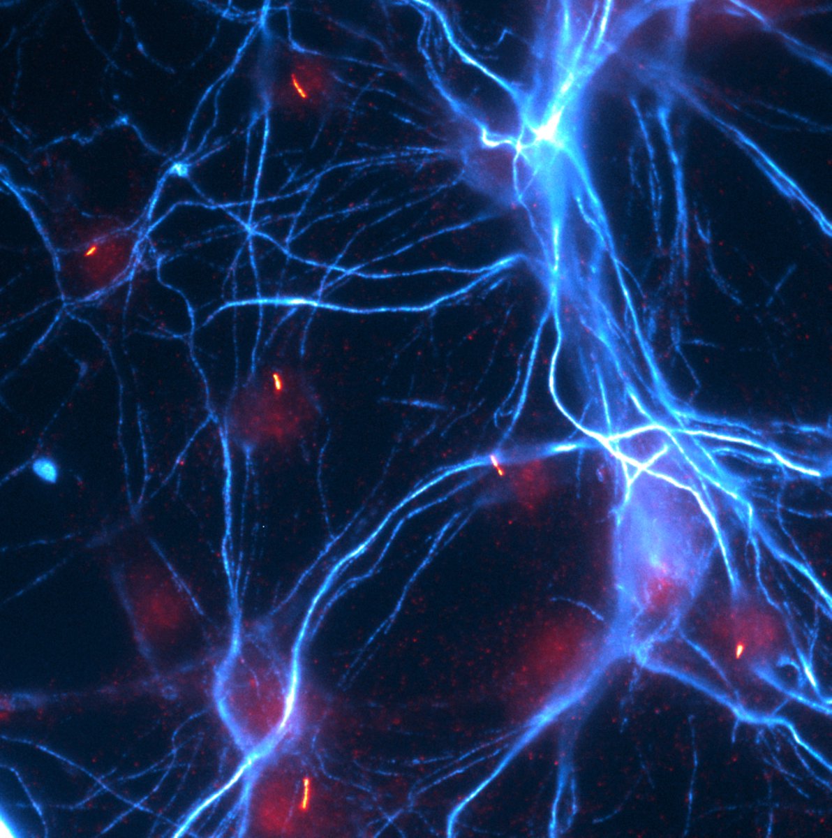
Chicken commissural neurons stained for neurofilament-M (Cyan) and the primary cilium marker Arl13b (red). happy #FluoresenceFriday !
4
36
233
Chicken commissural neurons stained for neurofilament-M (Cyan) and the primary cilium marker Arl13b (red). happy #FluoresenceFriday !
0
4
35
RT @Dev_journal: To find out more about this work, we caught up with first author, Alexandre Dumoulin, and corresponding author, Esther Sto….
0
1
0