
sudhir babu
@sudhirpath
Followers
48
Following
48
Media
12
Statuses
40
Dermpath/Renal Pathologist, Vijaya Diagnostic Centre
Vizag, India
Joined August 2018
RT @JZRenalPath: Capillary wall "eye-lashes" not always amyloid. Rare finding in a recent fibrillary case. IF = IgG. IHC = DNJAB9. Congo r….
0
29
0
RT @JZRenalPath: Older patient with anasarca. Bx with massive glomerular amyloidosis - AH type. IF and EM reveal concurrent heavy chain dep….
0
25
0
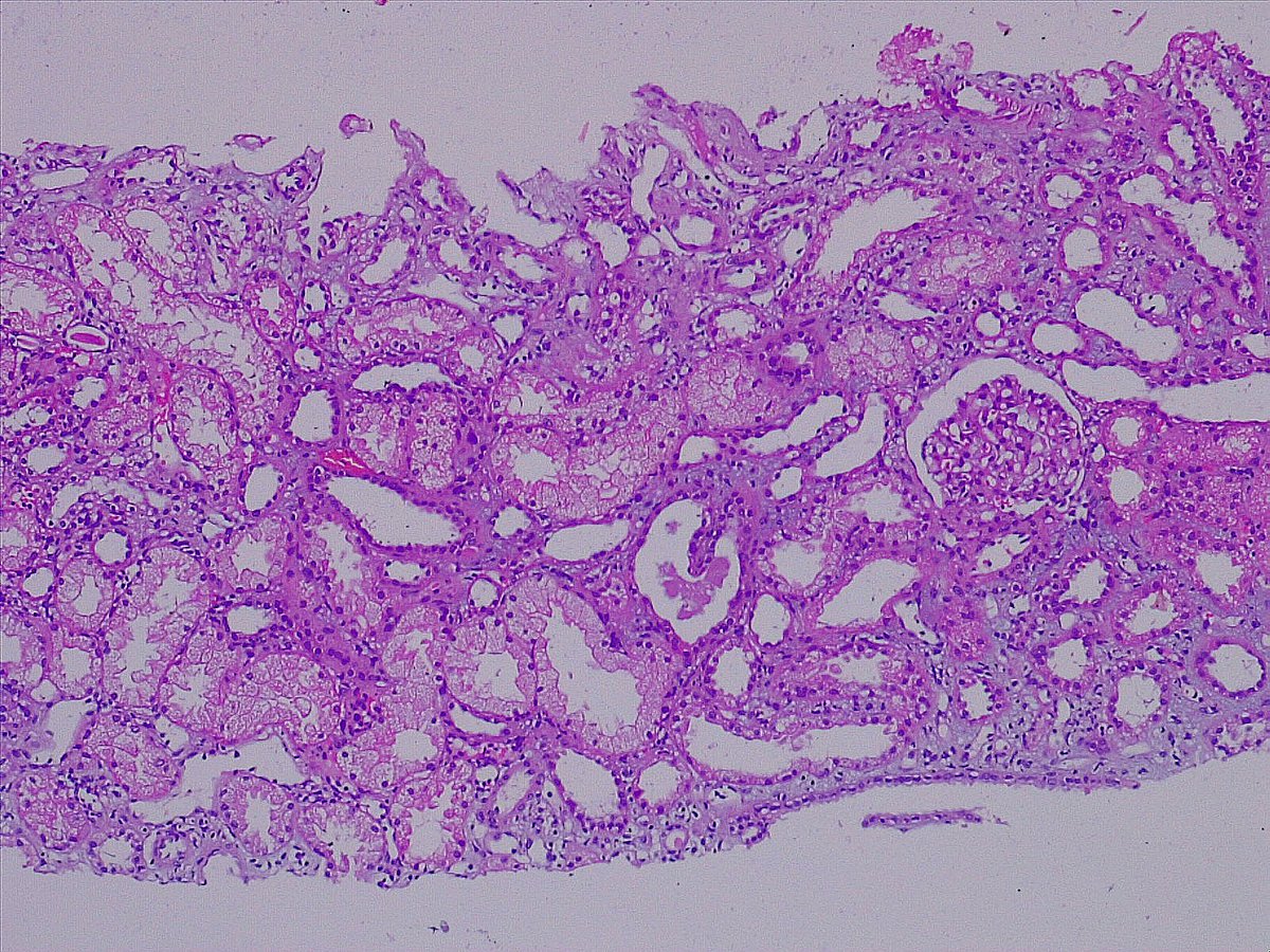
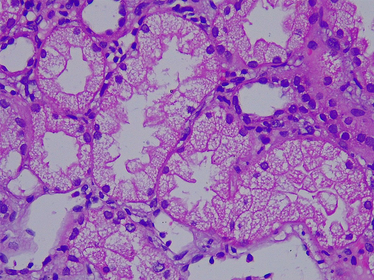
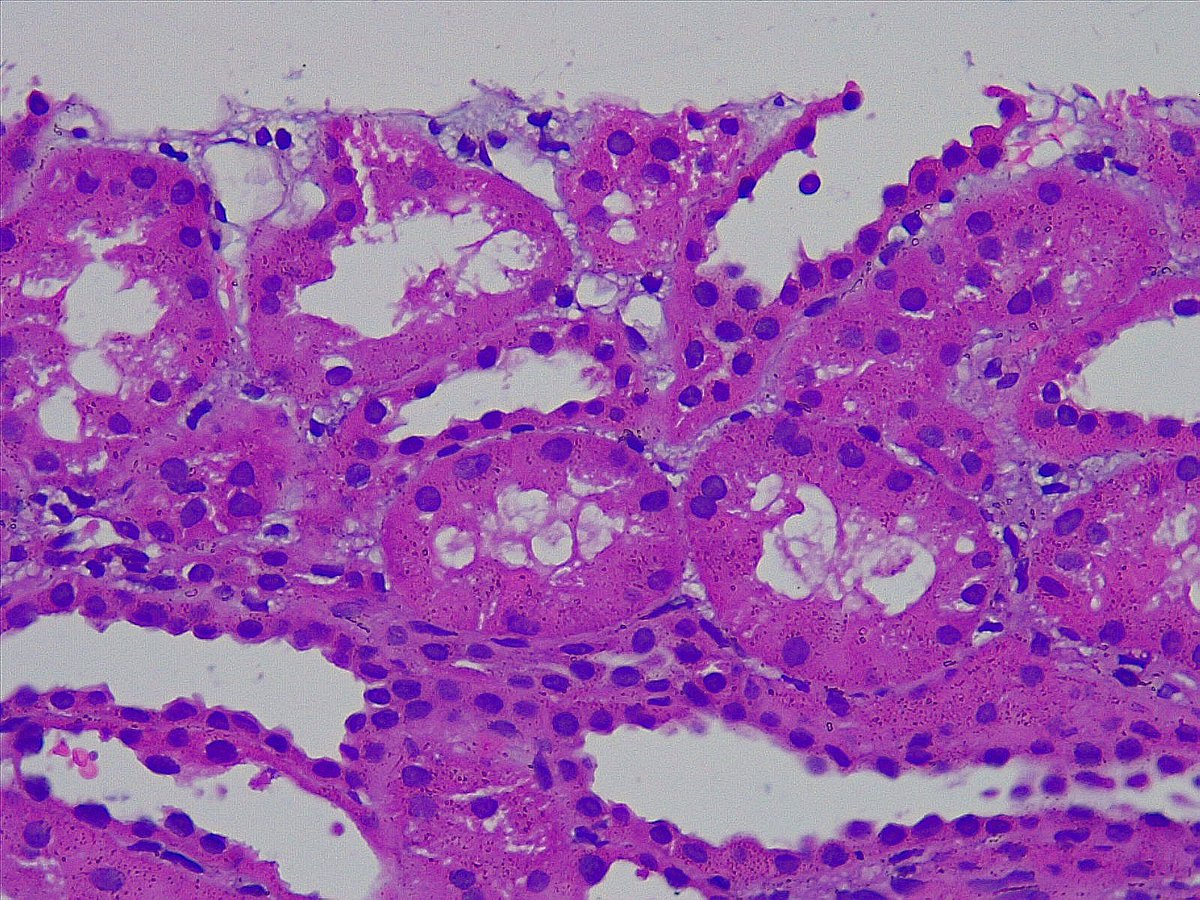
Glomerular foam cells occluding the capillary loops (Bloodless glomeruli) in a case of Acute CNI toxicity #renalpathology #CNItoxicity #isometricvacuolization.@draalok @RitambhraN @vinaypush @JZRenalPath.@Renalpathsoc


0
7
26
RT @JZRenalPath: Nice example of striped fibrosis which is often attributed to chronic CNI toxicity in kidney transplant biopsies. #renalpa….
0
58
0
RT @JZRenalPath: AKi in a pt with recent contrast administration and antibiotic use. Features of contrast nephropathy and allergic intersti….
0
22
0
@draalok @RitambhraN @vinaypush @JZRenalPath @Renalpathsoc Before making a diagnosis of acute CNI toxicity with Tubular foam cells, we need to think of: .1. Ischemic acute tubular injury.2. Osmotic tubulopathy.3. Potassium depletion.4. Nephrotic syndrome.
0
0
1
Isometric vacuolization of tubular epithelial cells (foamy tubular cells) in a case of Acute CNI toxicity #renalpathology #CNItoxicity #isometricvacuolization @draalok @RitambhraN @vinaypush @JZRenalPath @Renalpathsoc

2
16
34
RT @JZRenalPath: A reminder that cryoglobulinemic glomerulonephritis can still occur post-HCV treatment. Biopsy for hematuria, proteinuria,….
0
16
0
Finally, we got Kappa light chain restriction on direct immunofluorescence confirming the diagnosis of Light Chain Deposition Disease (LCDD)


0
0
4
Here comes the PAS stain, which shows PAS positive mesangial nodules ruling out amyloidosis.

2
0
4
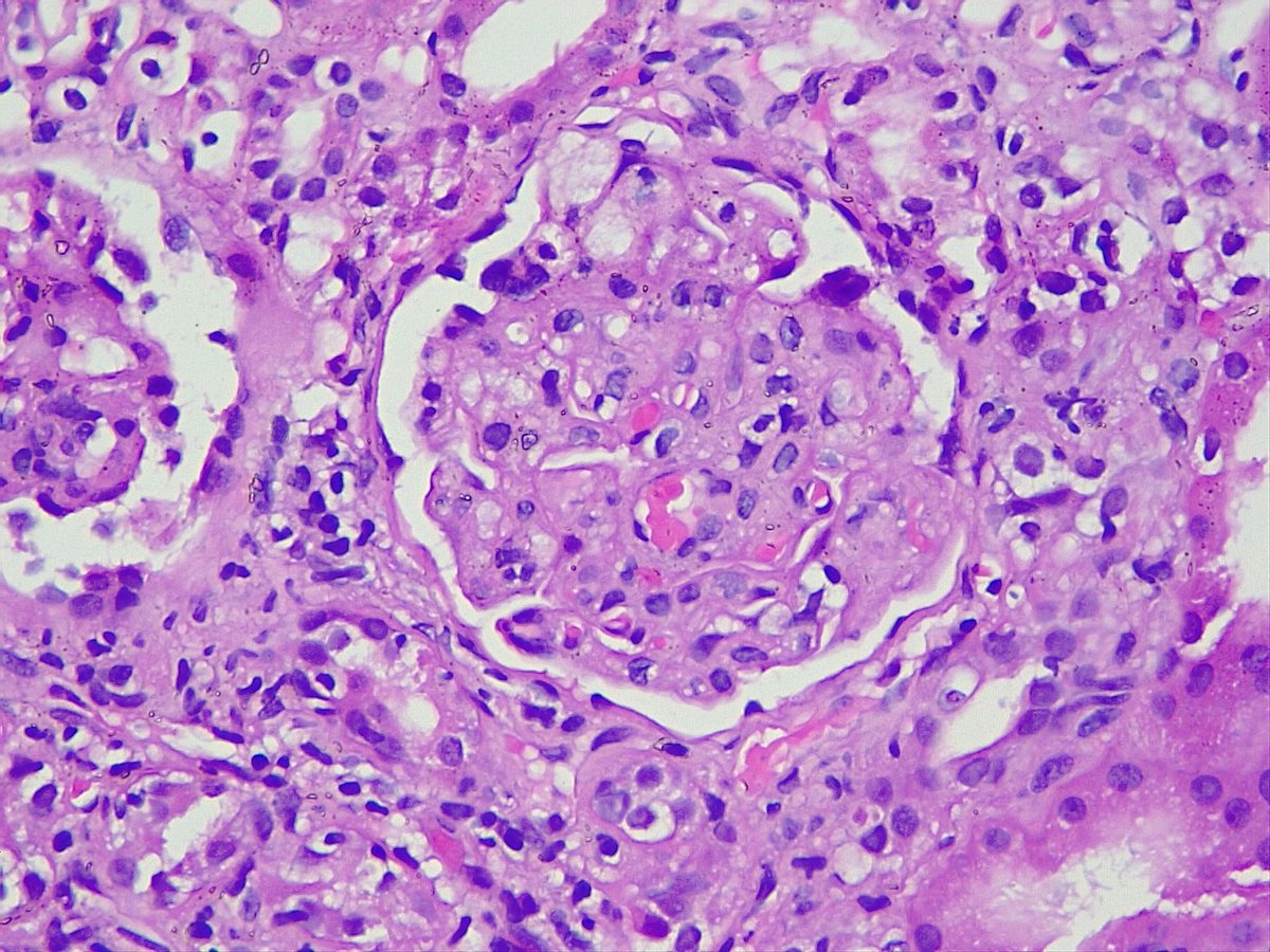
With this H&E picture, one should think of .1) Diabetic nephropathy (but the patient is non-diabetic).2) Idiopathic nodular glomerulosclerosis (but there is no smoking history and mesangial nodules are of different sizes).3) Amyloidosis.4) MIDD. 0.
2
0
3
Nodular glomerulosclerosis in a 48/M, with nephrotic range proteinuria, anemia, hypertension, chronic rheumatic heart disease #Renalpathology #mesangialnodules #nodularglomerulosclerosis @draalok @RitambhraN @vinaypush @JZRenalPath @SethiRenalPath @Renalpathsoc

1
13
30
Full House pattern is not specific for lupus nephritis. Check this article for more info 👉Wani AS, Zahir Z, Gupta A, Agrawal V. Clinicopathological Pattern of Non-lupus Full House Nephropathy. Indian J Nephrol.2020 Sep-Oct;30(5):301-306.
0
1
1
Ever wonder about the name 'FULL HOUSE'?.Just like Poker's 3 of a kind + a pair, .in Nephrology, its Ig (3 of a kind) + complements (a pair)!


2
0
5

Exploring the 'FULL HOUSE' pattern in Class IV Lupus nephritis! #Renalpathology #fullhousepattern #lupusnephritis #nephrology @DrRachnakhera @draalok @RitambhraN @vinaypush @JZRenalPath @SethiRenalPath @Renalpathsoc


1
5
8
In 1957, renal pathologist David Jones defined Membranous nephropathy, using methenamine silver stain to highlight GBM changes and spikes. As a result of his contributions, the Jones methenamine silver stain bears his name. #Medicalhistory #Nephrology.
0
0
2
GBM spikes on Jones methenamine silver stain in a case of Class III+V lupus nephritis. #Renalpathology #GBMspikes #lupusnephritis @DrRachnakhera @draalok @RitambhraN @vinaypush @JZRenalPath @SethiRenalPath @Renalpathsoc


1
2
13
@DrRachnakhera @draalok @SethiRenalPath @JZRenalPath @Renalpathsoc @PreethiSekarMD Spotting amyloid deposits: While the gold standard is Congo red stain under a polarizer microscope, a simpler approach is visualizing it under a fluorescent microscope. Enhance your diagnostic skills in amyloidosis. #Pathologytips #AmyloidRecongnition

0
0
1