Explore tweets tagged as #Adas3D

In a recent atrial flutter ablation in a 4-year-old patient with HLHS and Glenn circulation, CT images were pre-processed with ADAS 3D Medical for anatomical segmentation. This step allowed clearer visualization of the complex anatomy and supported precise procedural planning.
0
2
8
#ADAS3D LV Analysis from CT & MRI: ✅ Automatic segmentation of LV, LA, AO & LAA from CTA ✅ 3D visualization of fibrosis, to understand its distribution and transmurality ✅ Automatic detection of border zone 3D corridors ✅ LV wall thickness mapping from CTA ✅ Hypo-enhancement
0
1
13

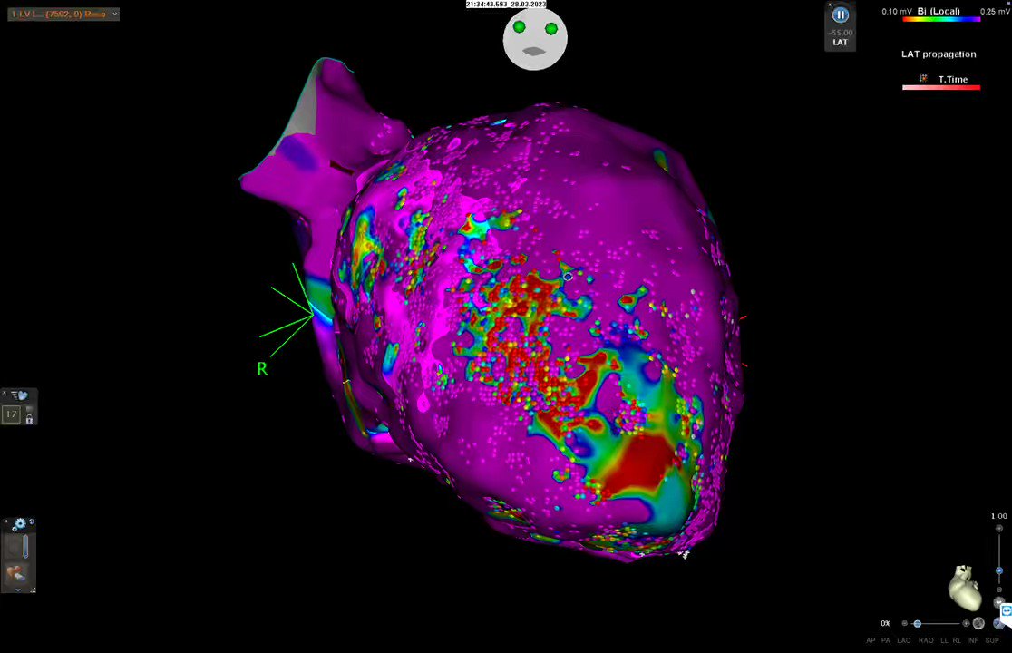
@FYang_EP @EffieNitsdoc @pjsm83 @aportasanchez @DhanjalTarv @ADAS3D @sozi81 @DrRoderickTung @mike_lean @Hapa_EP @Arritmias_HRC @ivroca @Callme_DrNeil @andresenriqueza NSR with LAM (0.1-0.25mv)

0
3
13

Absolutely outstanding workshops at Hospital Clínic de Barcelona with @ivroca @aportasanchez and team. During our stay we have also visited @ADAS3D office and hopefully will soon be able to implement it into our workflow. Thanks and hopefully see you soon ! #EPeeps

1
1
11
#ADAS3D LA Analysis from CT & MRI: ✅ Enhancement distribution mapped on LA surface ✅ Automatic detailed visualization of patient anatomy ✅ Visual maps of proximity to critical structures ✅ Identification of ablation gaps in LA From image to insight – supporting safer, more
0
0
5

@vish_luther Yes I have! Here is the @ADAS3D image (thanks @RosaMFiV) of the CT. The anterior area was around 2 mm thick!

2
5
18

Our current understanding of post-MI VT substrate derives heavily from completed MI model. We used a more contemporary reperfused MI swine model w/ 90min mid-LAD occlusion and studied scar with sequential LGE-CMR, @ADAS3D , functional substrate mapping & programmed stim. 2/5

1
3
11
https://t.co/DQPuyjmWlk Thank you Dr. @cortezdias, for this outstanding case of cardioneuroablation for recurrent vasovagal syncope at Hospital de Santa Maria, Lisbon — with #Adas3D CT fat pad reconstruction enabling precise anatomical targeting, tailored with extracardiac vagal
0
4
6
https://t.co/lyQyUIJ2Xo Imaging guidance opening new perspectives in CNA. Fascinating case, grateful to Dr. Procolo Marchese for sharing. The successful use of #ADAS3D analysis in a challenging cardioinhibitory syncope case highlights the promise of this approach. #Epeeps
0
0
0
The TRAMPOLINE VT study @NHSBartsHealth: combining Biosense Webster's OPTRELL catheter with MRI data from @ADAS3D to define culprit corridors within ventricular scar. With thanks to @jjmt_ep! #ablateVT #epeeps @chris_monkhouse @AlexGrimster @LambiasePier

1
16
45

Strong cooperation with Martin Eichenlaub, Amir Jadidi & Thomas Arentz🙏💪 @ivroca @ADAS3D @AG21_DGK @masitges @DGK_org @escardio @EduGuasch @tolosanaviu @arbelo_e @aportasanchez @AnaGarcalvarez2 @YoungDgk

0
2
7


The Power of #OpenEP ⚡🤝 -Sarcoidosis Patient with 2 VT’s -Septal Mid-Myocardial Scar and Substrate identified with ADAS 3D prior to case -Treated Robotically @MSMorningside @ADAS3D
#CanYourCatheterDoThis #EPeeps #RoboticEP


2
7
19


Incessant VT did not allow substrate mapping. HDgrid in CT channel @ADAS3D, diastolic EGM in <1 minute. RF at the exit of CT channel stopped VT in 10 sec. No more incessant V and full substrate ablation was completed. #AblateVT @hospitalclinic Imaging+HD mapping working together
1
28
131

“ AWMI -VT “….@ADAS3D - cardiac magnetic Resonance-aided VT substrate mapping and ablation ,at Madras Medical Mission , chennai Tx @UlhasDr @anindya_0211
@AbbottCardio @murphy_lzy @Yogesh_EP @dineshkrishn @Harishs1794 @RYeddanapudi @rahul_bme53

1
14
51


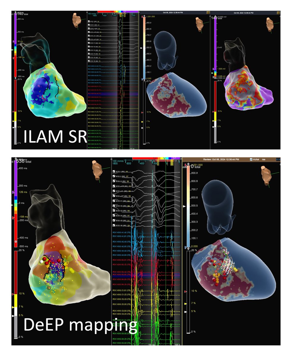
cMRI-aided VT ablation. Perfect match of MRI corridors with isochronal late activation maps (DeeP mapping). Minimal lesions with short procedural time (90 min). @SteliosDragasis @athsaplaouras @ipetemil
@Kariki_O @BellosPeriklis @eva_nyktari
@MEfraimidis @ADAS3D OCSC
1
25
71