
Yale Biomedical Imaging Institute
@YaleBioImaging
Followers
11
Following
12
Media
8
Statuses
28
Yale BioImaging is an interdisciplinary research institute transforming our understanding of health and disease through biomedical imaging
New Haven, CT
Joined January 2025
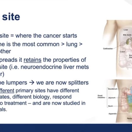
Director Georges El Fakhri presented on imaging approaches for #NETS Awareness Day 2025. See link for a full video of the Smilow Shares seminar, highlighting the diagnosis, treatment, and awareness of neuroendocrine tumors:
medicine.yale.edu
On November 10, 2025, Dr. Pamela Kunz hosted a Smilow Shares in honor of NETS Awareness Day.
0
0
0
Our finance and operations team was recently honored with the Lorimer Award, which celebrates outstanding Yale staff. This team worked tirelessly to provide the financial and operational expertise needed to launch our new Yale Biomedical Imaging Institute. https://t.co/tQPOmnIkEo
0
0
0
Two of our institute members have been recognized for their highly cited research by the data and analytics company Clarivate: John Krystal, @DScheinost
https://t.co/WAIKAKuazj
medicine.yale.edu
Yale School of Medicine faculty members were recognized for publishing studies that rank in the top 1% based on the number of citations they received in their
0
0
0
Congratulations to Prof. Jason Cai for his startup "Synvest Imaging Inc.: Developing novel molecular imaging agents to advance early detection and personalized treatment".

The 2025 Yale Faculty Innovation Awards honor academic founders whose startups—rooted in Yale research—are advancing breakthroughs in health, sustainability, and engineering. Meet the awardees: https://t.co/TUHsv7FKXk
0
1
0
The 3rd annual Nonlinear Parameter Parley and Pub Crawl, held at Seoul National University Hospital on May 30, brought together PET kinetic modeling experts. New topics included time-varying models & spatial drug occupancy #NLPPPC #PETImaging #BrainPET2025
https://t.co/UiR4dgRCP9
medicine.yale.edu
The third Nonlinear Parameter Parley (NLPP 3.0) was held on May 30, 2025, at Seoul National University Hospital. Organized by Dr. Su Jin Kim, Prof. Evan Morris,
0
0
0
Like a caricature artist does with facial features, Yale Department of Radiology and Biomedical Imaging’s @DScheinost, MD, and colleagues are emphasizing distinctive features of individuals’ brain activity to better predict their behavior: https://t.co/WhvyUUsnCl
@YaleRadiology
medicine.yale.edu
Emphasizing individual differences in brain activity data improves predictions of several characteristics and behaviors.
0
3
5

Resting-state FC has long been a source of debate. Recent evidence suggests that task-like patterns dominate its signal. To study less dominant components, we introduced a caricaturing method to project the data to a subspace orthogonal to these patterns. https://t.co/B5S7xxReOk
1
3
2
YBII members present a method in @NatureNeuro to remove task-like signals from resting-state fMRI, boosting individual differentiation and fingerprinting accuracy. Their work reveals overlooked signals beyond dominant co-activation patterns. https://t.co/GgWpr9VSgj
0
1
1
🚨 New paper alert! 🧠 Head motion during brain PET scans causes artifacts & quantitative errors but hardware tracking isn’t always feasible! 🔥 Discover DL-HMC++: a deep learning model that predicts motion directly from PET raw data! 🔗 https://t.co/GS5v4z80NQ
0
2
5

Carotid artery image-derived blood time–activity curve (CA ID-BTAC) extraction with minimal bias is feasible with ultra-high-resolution brain-dedicated PET scanners. https://t.co/r7Dkyb9Dde
#NuclearMedicine #MolecularImaging #PETscan @YaleMed @tommaso_volpi
0
4
11
Dr. Jason Johnson and collaborators developed the world’s first DL model specifically designed to segment ultra-high resolution brain MRI. The new GA-MS-#UNet++ model uses advanced attention mechanisms to unlock the potential to detect subtle brain changes https://t.co/dctzavIorH
0
1
2

First official Bini lab preprint is live! https://t.co/n2MJ67VOxp Many thanks to @Faraz_Nejati @FrnkEbrahimian Rui Ren and Yuan Huang for their hard work on this study! @YaleBioImaging
medrxiv.org
Objective To determine if combining PET-derived beta-cell mass (BCM) estimates with MRI- based morphology metrics improves the prediction of beta-cell functional mass in type 2 diabetes (T2D)....
0
3
10

Multiangle data acquisition and AI enhanced mapping to multiangle data both enhance image resolution in cardiac CZT SPECT perfusion imaging. Promising for improved diagnostic accuracy! @YaleMed @YaleCardiology #cvnuc Read now👉 https://t.co/q7BqlAmKNe
0
8
18
MICCAI workshops: Deep Generative Models Workshop (DGM4MICCAI): Keynote: Julia Wolleb https://t.co/hvYBW0FnsN
@YaleRadiology @YaleBIDS @YaleBme @YaleEngineering @YaleMed
0
0
1
MICCAI workshops: Advances in Simplifying Medical UltraSound (ASMUS): VidFuncta: Towards Generalizable Neural Representations for Ultrasound Videos Julia Wolleb, Florentin Bieder, Paul Friedrich, Hemant D. Tagare, Xenophon Papademetris https://t.co/fiRmV7783l
1
0
0
MICCAI: Adapting Vision Foundation Models for Real-time Ultrasound Image Segmentation Xiaoran Zhang, Eric Z. Chen, Lin Zhao, Xiao Chen, Yikang Liu, Boris Maihe, James S. Duncan, Terrence Chen, and Shanhui Sun https://t.co/ho0n3bh2CQ
1
0
0
Check out YBII at @MICCAI_Society!! MICCAI: Geometry-Guided Local Alignment for Multi-View Visual Language Pre-Training in Mammography Yuexi Du, Lihui Chen, Nicha C. Dvornek https://t.co/6np5UE7e6X
1
0
0
This developmental trajectory is consistent across four datasets, with deviations from this trajectory associated with poorer executive function. Girls also peaked earlier than boys. @Yale_INP @YaleBioImaging @YaleRadiology @YaleMRRCneuro
https://t.co/nCEKHjjbgT
0
1
1
Excited to share @JeanSYe_ work now out in @NeuroCellPress! We found that during development, people become more variable in how much they engage recurring brain states.
1
4
7
A new #PET tracer can provide insights into how spinal cord injuries affect not only the spinal cord, but also the brain, according to our new research published in @JournalofNucMed
https://t.co/6MuCXe7NjO
0
1
2