
Tom Sheard
@TMDSheard
Followers
701
Following
5K
Media
612
Statuses
3K
No more posting on this account... Find me on Linkedin or Instagram.
Sheffield, England
Joined November 2013
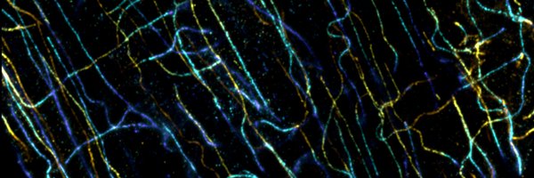
Are you a microscopist/biologist seeking new ways to visualise subcellular compartments? 🔬Our new paper in @nanoscale_rsc shows how fluorescent esters can give you valuable morphological insights into cellular architecture 🦠(1/6)

5
39
191
Excited to announce I've joined @UKNikon as an Advanced Imaging Specialist! 🔬 Helping the North + Midlands to harness Nikon's portfolio of microscope systems. Looking forward to continuing my work in the amazing microscopy community. Reach out to chat about advanced imaging!

5
2
68
Huge thanks to all my amazing labmates over the years (you know who you are!), and to my super collaborators across Biosciences, Edinburgh, Leeds, Oxford, Italy, Denmark and New Zealand. And big thanks to the facility staff who made the science possible! ✨🔬.
0
0
2
I'd like to express my deepest gratitude to Izzy @i_jayas, the kindest mentor anyone could ask for. It's been an absolute pleasure working with you over nearly 8 years since starting the PhD in Leeds!.
1
0
1
After 3 wonderful years in @mcb_sheffield, my postdoc journey comes to an end. I've loved my time in Firth Court, thanks to a lot of brilliant and friendly people. Onto the next exciting chapter in July (more on that later👀). Now for a sunny holiday!

5
1
57
Really enjoyed my first ever #Elmi2024 in Liverpool! Great week capped off with dinner + a good ol' boogie to live band at @TitanicHotelLiv . Many thanks to the organisers + volunteers! @RoyalMicroSoc


0
2
20

Strong finish from team academia, tactical decision by @YorkBioimaging to go for the 4-5-7 formation paid off! #elmi2024

1
1
16
Oh @Tool. No words do this band or watching them live justice. Forever my favourite band of all time.

3
1
12
RT @stephan_till: After countless requests, we're thrilled to introduce PK Mito Orange FX - a fixable variant of PKMO. PKMO FX allows immun….
0
71
0
Pleased to hear our recent paper has been included in the @nanoscale_rsc Most Popular 2023 Articles collection!
Are you a microscopist/biologist seeking new ways to visualise subcellular compartments? 🔬Our new paper in @nanoscale_rsc shows how fluorescent esters can give you valuable morphological insights into cellular architecture 🦠(1/6)
0
4
20
Time to find another note-taking app, any suggestions? Used @evernote for 5 years, made 1000 notes(!) to help me complete PhD + postdoc projects, but this is the end! 😢 Free limit of 50 notes is absurd, twinned with terrible support and lacklustre user experience. Seeyaa! 👋.
5
0
0
Plate approach for expansion microscopy. Crucially making it easier to do pre- post- expansion imaging for error identification. Also streamlining process to reduce manual handling of gels + increasing throughput. Nice work @rajseehra + rest of our team!.
0
3
14
Another day, another incredible expansion microscopy paper!.
Its out !!! 🥳. Very proud to announce that my PhD work is now published in Nature communication: We developed #iUExM, an optimized iterative expansion protocol allowing 16 to 26 fold homogeneous expansion together with specific bright labelling !

0
1
10
Looking forward to connecting with other microscopists doing exciting science at the upcoming Super-resolution in the North meeting in York🔬 Registration is free! #SRITN2023 @RoyalMicroSoc.

Announcing Super Res In the North in York on 15 Dec! Three reasons to register now: .✅ Awesome line-up @seamus_holden @ESRIC_imaging @AleksPonjavic &c!.✅ FREE both in-person and virtual.✅ Selected early-career flash talks open!. (1/2)

0
2
6
Many thanks to the co-authors @i_jayas @kinsuen__ @t_shakespeare @rajseehra and Michael Spencer, team in @mcb_sheffield imaging facility🔬, and the funders IBIN and UKRI who made this research possible! Blog post for the new paper can be found here: (6/6).
1
0
2
Esters are super useful in many samples (monolayers + multi-cellular specimens - eg C. elegans . For other examples of esters as imaging probes, check out amazing papers - panExM (@OnsMSaad1), wholeExM (@jaebyumchang) and #ONE_microscopy (@AliHShaib) (5/6).
life-science-alliance.org
Correction to Materials and Methods.
1
0
2
We also report new intricacies in labelling patterns which arise when multiple esters are added together, through multi-ester interactions (involving FRET between the dye molecules), and complexities which also depend on the timepoint of ester addition. (4/6)

1
0
1
Esters are useful for visualising overall morphology, but are perhaps most powerful as a counterstain alongside specific labels. We show how esters used in tandem with immunolabelling can give valuable contextual info, revealing targets in relation to cell architecture. (3/6)

1
0
5
In the paper you can find catalogues of images for 14 esters conjugated to a bunch of dyes 🔆 (eg Alexa, BODIPY and ATTO), with a range of charge + hydrophilic properties. We used 4x expansion microscopy but esters can also provide insight for non-super-res experiments too! (2/6)

1
0
3