
Int Soc Dermpath
@IntSocDermpath
Followers
3K
Following
1K
Media
597
Statuses
2K
Half Moon Bay, CA
Joined November 2016
1st European Joint Meeting of the International Society of Dermatopathology #ISDP #dermatopathology Dr. Kempf on CD8 lymphoproliferative disorders
0
5
27
1st European Joint Meeting of the International Society of Dermatopathology program #ISDP #dermatopathology
0
1
4
1st European Joint Meeting of the International Society of Dermatopathology an European Society of Pathology starts today in Vienna. First talk is by Dr. Fraitag #ISDP #dermatopathology
1
4
20
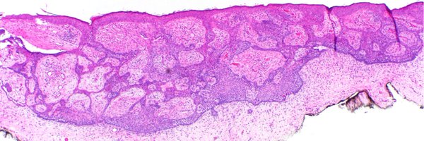
#ISDP #Olympics Spot diagnosis Case 20 - @JCandidoXavier CHROMOBLASTOMYCOSIS •Pigmented (dematiaceous) fungi •Prominent pseudoepitheliomatous hyperplasia •Granulomatous infiltrates with round brownish Medlar bodies resembling copper pennies or coffee beans #path #derm
0
11
33
#ISDP #Olympics Spot diagnosis Case 19 - @JoyceSSLee SEBACEOUS CARCINOMA • Periocular (75%) • Basophilic germinative sebaceous cells • Sebocytes with vacuolated cytoplasm • Nuclear pleomorphism • Increased and atypical mitoses • Necrosis #Path #derm #pathology
2
11
36
#ISDP #OlympicGames Spot diagnosis Case 18 - @JCandidoXavier SYRINGOCYSTADENOMA PAPILLIFERUM •It can occur in naevus sebaceus of Jadassohn •Endophytic crateriform lesion •Two layers of cells forming papillary and cystic structures • Numerous plasma cells #path #derm
0
5
21
#ISDP #OlympicGames Spot diagnosis Case 17 - @JoyceSSLee CUTANEOUS ROSAI DORFMAN DISEASE •Non-Langerhans cell histiocytosis •Large histiocytes exhibiting emperipolesis (containing inflammatory cells) •CD68+, S100+, CD1a- •Lymphoplasmacytic infiltrate #Path #derm
0
12
28
#ISDP #Olympics Spot diagnosis Case 16 - @JCandidoXavier ELASTOFIBROMA •Benign soft tissue tumor characterized by abnormal elastic fibres and fibroblasts •Hypocellular tumor with fragmented and thickened to globular elastic fibres #Pathology #path #dermatology #derm
0
5
21
#ISDP #OlympicGames Spot diagnosis Case 15 - @JoyceSSLee PLEVA: •Parakeratosis •Apoptotic keratinocytes •Basal vacuolar alteration •Dermal and intraepidermal hemorrhage •Wedge-shaped lymphohistiocytic infiltrate •+/- Lymphocytic vasculitis #derm #path #pathology
0
5
16
#ISDP #OlympicGames Spot diagnosis Case 14 - @JCandidoXavier PARACOCCIDIOIDOMYCOSIS •Dimorphic fungi (mycelial in the environment and yeast in the host) •Granulomatous reaction pattern •Round yeast with single or multiple buds with narrow necks. #derm #path #pathology
1
4
20
#ISDP #OlympicGames Spot diagnosis Case 13 - @JoyceSSLee ONYCHOMATRICOMA •Thickened nail plate •Wormwood-like or honeycomb-like cavities •Papillary digitate projections covered by matrix epithelium •V-shaped keratogenouszones •Fibrocellular stroma #path #dermatology
2
6
18
#ISDP #OlympicGames Spot diagnosis Case 12 - @JCandidoXavier LANGERHANS CELL HISTIOCYTOSIS • Round/ovoid medium-sized cells with indented, lobulated or coffee-bean nuclei. • Abundant and eosinophilic cytoplasm •IHQ markers: CD1a, Langerin and S100 protein #derm #path
1
0
9
Join us for three days in Queenstown, New Zealand for insightful discussions, engaging presentations, and networking opportunities with leading experts and professionals in the field of Dermatopathology. #ISDP #Dermpath #path #pathology
@JMGardnerMD @GeronimoJrLapac
0
9
17
#ISDP #Olympics Spot diagnosis Case 11 - @JoyceSSLee NECROBIOTIC XANTHOGRANULOMA •Areas of necrobiosis •Xanthogranulomatousinflammation (foamy histiocytes, Touton giant cells) •Cholesterol clefts •Large, bizarre angulated giant cells with many nuclei #PathTwitter #derm
1
0
12
#ISDP #Olympics2024 Spot diagnosis Case 9 - @JoyceSSLee NECROTIZING INFUNDIBULAR CRYSTALLINE FOLLICULITIS •Folliculocentric crater •Eosinophilic filamentous material •Material likely derived from Malassezia yeasts, bacteria, and/or sebaceous lipids #path #dermatology
1
10
31
#ISDP #Olympics Spot diagnosis Case 8 - @JCandidoXavier OCHRONOSIS •Clinical presentation: blue–black cutaneous pigmentation •Typical swollen, irregular, golden brown banana 🍌shaped, sickled or round ochronotic bodies #dermpath #dermatology #pathology #path
1
2
16
#ISDP #Olympics Spot diagnosis Case 7 - @JoyceSSLee HAND, FOOT AND MOUTH DISEASE •Caused by coxsackievirus A16, enterovirus 71 •Intraepidermal blister •Ballooning and reticular degeneration •Epidermal necrosis •Neutrophils #Dermatology #derm #dempath #pathology
2
9
24
#ISDP #olympics Spot diagnosis Case 6 - @JCandidoXavier GRANULAR CELL TUMOR •Benign neuroectodermal tumor •Large cells with abundant and granular cytoplasm •Synonym: Abrikossoff tumor •Pseudoepitheliomatous hyperplasia can be prominent #path #pathology #dermatology
1
6
25
#ISDP #Olympics Spot diagnosis Case 5 - @JoyceSSLee SCLEROMYXEDEMA •Haphazard proliferation of fibroblasts within the reticular dermis •Fibrosis and skin thickening •Increased dermal mucin #dermatology #dermatopatology #dermapath #path
1
2
16