Head and Neck Radiology
@HeadNeckRads
Followers
12K
Following
699
Media
285
Statuses
2K
Learn with me about imaging above the clavicles, especially from dura to pleura. #HNRad #NeuroRad #RadEd Curated by @francisdeng
Joined October 2019
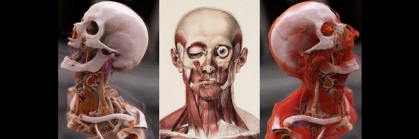
RT @daniel_gewolb: ⭐️What is the most likely diagnosis in this patient presenting with a lump on the head?. More images in 🧵. #MedEd #medic….
0
14
0
History: Hoarseness and cough.Finding: Severe interarytenoid edema.Diagnosis: Laryngopharygeal reflux.Treatment: Acid suppression


1
14
101

Generate videos in just a few seconds. Try Grok Imagine, free for a limited time.
416
688
3K
RT @jussihirvonen: Thrilled to share the @ESHNRSociety practice recommendations for acute head and neck infections, published in the @myESR….
link.springer.com
European Radiology - Acute head and neck infections are common in the population and can have serious complications. Prompt diagnosis and treatment are necessary to avoid morbidity and mortality....
0
19
0
RT @PhilipRChapman1: Now is the time to register! . Excitement is building for the 59th Annual Meeting of the American Society of Head and….
0
14
0
RT @DocNavarrow: ✅Not all parapharyngeal tumors are salivary, neurogenic or paraganglionic. 💡If you see skull base extension and vascular….
0
25
0
RT @daniel_gewolb: 🔷75 y/o M with progressive left face discomfort and enlargement over 2 weeks following dental extraction. What is your d….
0
14
0

RT @LyellJ: Neurology is so cool:. A 61 year old man comes in for another opinion on his diagnosis of ALS. Several years of progressive upp….
0
127
0

RT @DocNavarrow: 🤷♂️A 23-year-old woman presents with a painless, progressive swelling in the left submandibular region and an intraoral b….
0
13
0
RT @piandro3142: Important from @francisdeng #Radiopaedia2025 re: neurovascular compression of CN VII - from the root exit point to the pro….
0
22
0
RT @journalneurooph: Point Counter-Point - Brain MRI Should Be Routinely Ordered in Patients Presenting With Acute Retinal Artery Ischemia:….
0
6
0
RT @francisdeng: Shock-like/stabbing throat, jaw, and/or ear pain on the left. CISS-MRA fusion image showing left PICA compressing CN IX ro….
0
10
0
Answers:.Case 1: Case 2: Both courtesy of Mostafa Elfeky.
0
0
2
TWO cases, similar shape and borders, different location. Can you name them?. From @DrAndrewDixon's cystic neck lesion talk @Radiopaedia Day 1


1
9
70
When reading H&N cancer post-treatment MRIs, look for cortical bone discontinuity that suggests osteoradionecrosis. This is harder than on CT, so you have to remember to look and distinguish this from simple marrow signal heterogeneity. @CMGlastonbury at @Radiopaedia Day 1

0
9
40
Radiologic foundations teacher extraordinaire @radiologytuts reviews the deep neck spaces on @radiopaedia Day 1. If you liked Michael Nel's physics lessons, you can look forward to the anatomy lecture library he's building now.


0
12
73
18F-fluorocholine PET shows a lot of promise in localizing parathyroid lesions. Look at that beautiful resolution and contrast! Makes sestamibi look ancient. Case from A Ragavan @Radiopaedia Day 1 with @DrAndrewDixon @SalAyesa @CMGlastonbury


0
1
7
On 4D CT for parathyroid, describe the superior-inferior level of parathyroid lesions according to anterior landmarks (eg, cricoid, thyroid isthmus and poles). Describing it by vertebral level is useless for the surgeon!.@Radiopaedia Day 1 with @DrAndrewDixon @CMGlastonbury

0
2
16
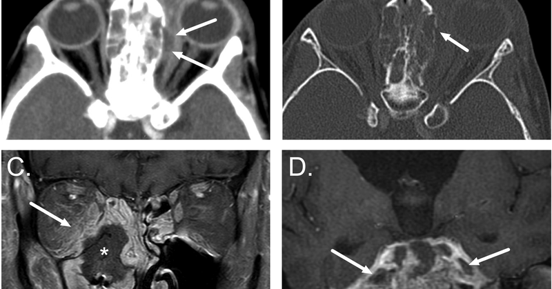
RT @drSurjthVattoth: Imaging Pearl 15: Congenital nasal pyriform aperture stenosis (CNPAS): Anatomy of PYRIFORM APERTURE of Nose & what all….
0
28
0