

Dr Navarro
@DocNavarrow
Followers
3K
Following
9K
Media
661
Statuses
4K
Neuroradiologist. MD, PhD, EDiNR. Academic Editor at European Journal of Cancer Care Advisory Editorial Committee, @RevistaRADIOLO2 No medical advice.
HGUC, Castellón
Joined August 2022
⁉️Are reference values in phase-contrast CSF flow studies truly reproducible? 📷I explore this question in my latest paper published in Neuroradiology: @NRADjournal #hydrocephalus.

1
5
17

RT @Fnmr_radiologue: 🚨 L'attaque est frontale ! . 📚 Les deux rapports « Charges et Produits » de la CNAM et « Pertinence en radiologie »….
0
9
0
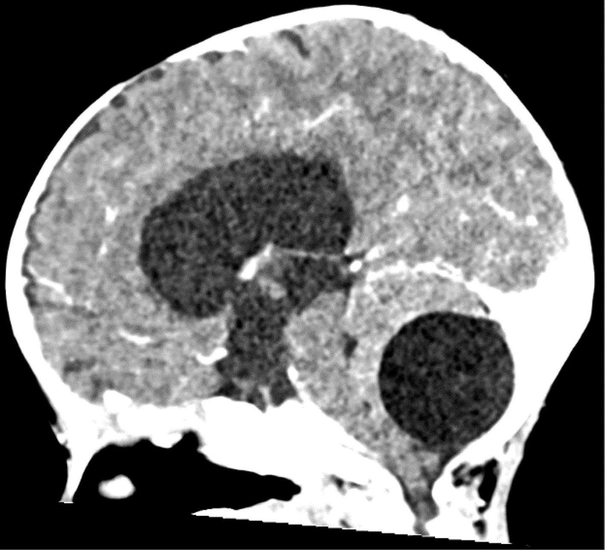
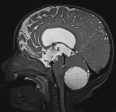
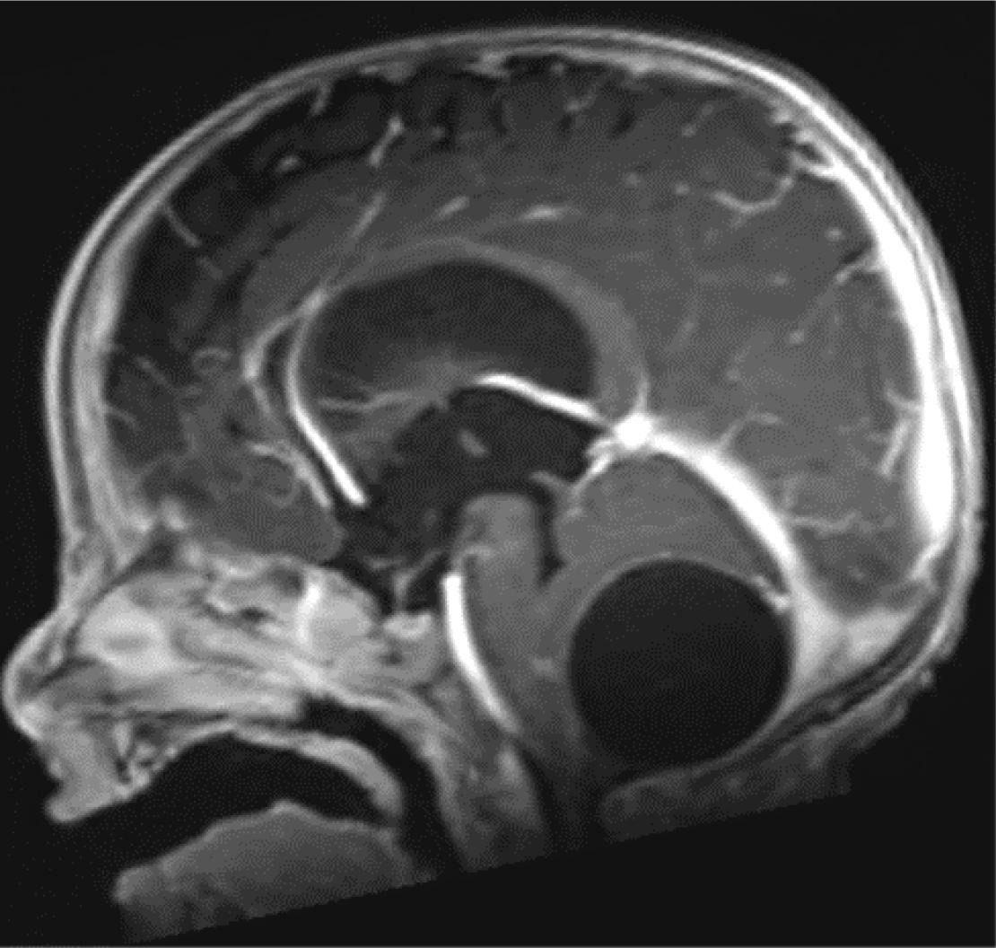
✅Mature cystic teratoma. In infants, posterior fossa cystic masses often suggest pilocytic astrocytoma, but absence of mural nodule, enhancement or solid component should prompt reconsideration. Teratoma may mimic benign cysts, even without fat.
👶A 6-month-old boy is referred due to increasing head circumference. Neurological exam is unremarkable. Serum AFP and β-HCG are negative. Surgery is performed the following day. Histology confirms the diagnosis. 🕵️♂️⁉️What would you suspect?

1
10
69
✅Mature cystic teratoma. In infants, posterior fossa cystic masses often suggest pilocytic astrocytoma, but absence of mural nodule, enhancement or solid component should prompt reconsideration. Teratoma may mimic benign cysts, even without fat.

0
2
7
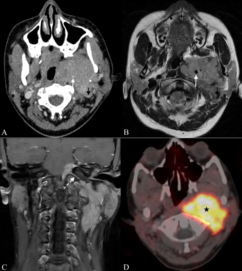
✅Not all parapharyngeal tumors are salivary, neurogenic or paraganglionic. 💡If you see skull base extension and vascular encasement, consider extracranial meningioma even in young adults.
🤷♂️A 23-year-old woman presents with a painless, progressive swelling in the left submandibular region and an intraoral bulge. No dysphagia, voice changes, or neurological deficits. 👨⚕️What diagnosis would you consider for this deep neck mass?
1
24
98
✅Not all parapharyngeal tumors are salivary, neurogenic or paraganglionic. 💡If you see skull base extension and vascular encasement, consider extracranial meningioma even in young adults.
0
0
12
👶A 6-month-old boy is referred due to increasing head circumference. Neurological exam is unremarkable. Serum AFP and β-HCG are negative. Surgery is performed the following day. Histology confirms the diagnosis. 🕵️♂️⁉️What would you suspect?
4
9
64
RT @DocNavarrow: 🤷♂️A 23-year-old woman presents with a painless, progressive swelling in the left submandibular region and an intraoral b….
0
13
0
🤷♂️A 23-year-old woman presents with a painless, progressive swelling in the left submandibular region and an intraoral bulge. No dysphagia, voice changes, or neurological deficits. 👨⚕️What diagnosis would you consider for this deep neck mass?
4
13
104
💡In multiple sclerosis, depression is linked to lesions in the raphe nuclei and locus coeruleus, key brainstem hubs of the serotonin and noradrenaline systems. ☹️MRI may help identify patients at risk before symptoms emerge.

2
12
76
✅ Erdheim-Chester disease. Consider ECD in the differential diagnosis of atypical enhancing brain lesions when lesions are bilateral, periventricular, or multifocal, especially if systemic findings are present.

ajnr.org
SUMMARY: Erdheim-Chester disease (ECD) is a rare, multisystem histiocytic disorder characterized by its variable clinical presentations. CNS involvement is observed in approximately one-half of...
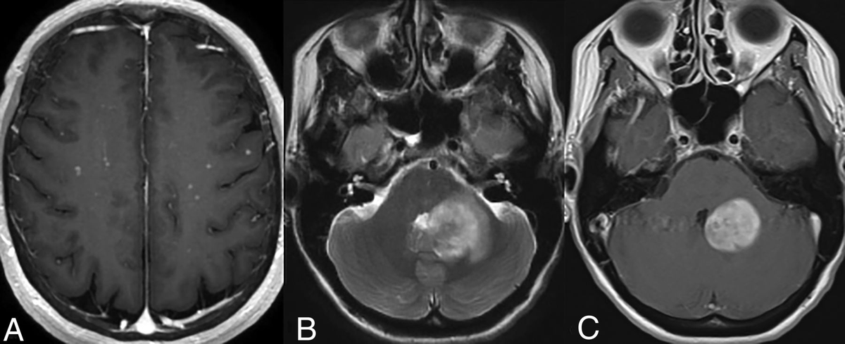
🧔♂️One patient has scattered bilateral micronodular enhancing lesions. 👨🦲Another shows a periventricular T2-hyperintense masslike lesion with homogeneous enhancement and mild edema. Same diagnosis in both. 🤔What disease are we dealing with?
1
24
104
✅ Erdheim-Chester disease. Consider ECD in the differential diagnosis of atypical enhancing brain lesions when lesions are bilateral, periventricular, or multifocal, especially if systemic findings are present.

1
2
12
RT @DocNavarrow: 🧔♂️One patient has scattered bilateral micronodular enhancing lesions. 👨🦲Another shows a periventricular T2-hyperintens….
0
12
0
🧔♂️One patient has scattered bilateral micronodular enhancing lesions. 👨🦲Another shows a periventricular T2-hyperintense masslike lesion with homogeneous enhancement and mild edema. Same diagnosis in both. 🤔What disease are we dealing with?
4
12
77
RT @francisdeng: The Radiopaedia 2025 conference is soon, July 21-25. Trainee or specialist, you’re going to learn something (w/ CME credit….
0
19
0
RT @pnavalbaudin: 🎥 Missed our Radiomics & Advanced Neuroimaging in Neuro-oncology webinar? The recording is now FREE: .
0
11
0
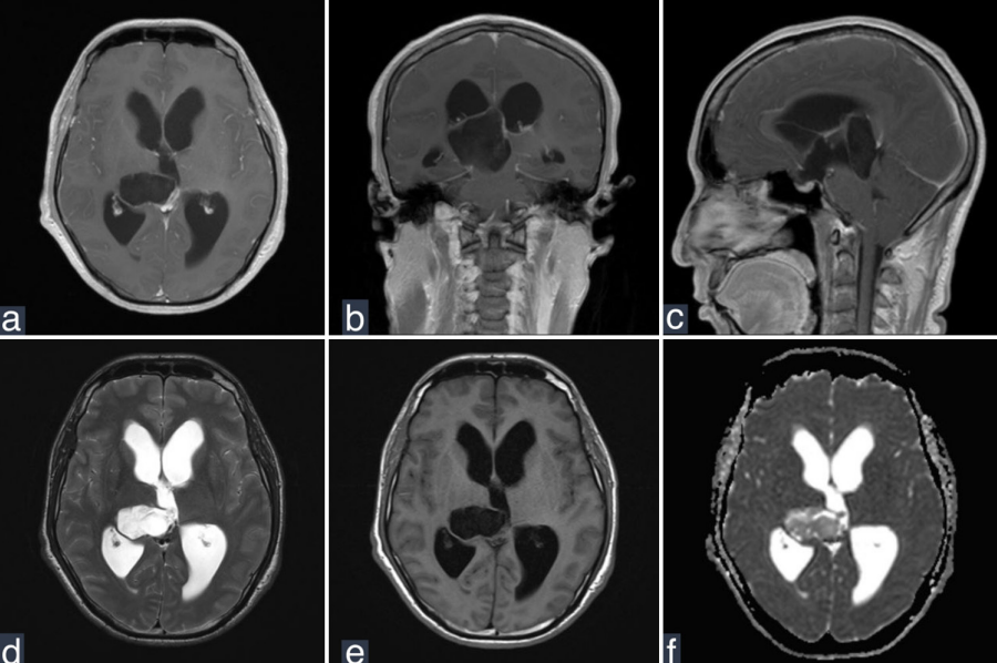
🤯Answer: Cholesteatoma — in the thalamus!. Yes, intraaxial and mimicking glioma on MRI. Only the second reported case worldwide. A pearly white mass, keratin-filled, deep in the brain. Not all that glitters is glioma.
surgicalneurologyint.com
🎙️NeuroQuiz Time!🧠. With the letter C. a rare intraaxial tumor that looks like a glioma on imaging but isn’t. What's your guess?
4
18
83
🤯Answer: Cholesteatoma — in the thalamus!. Yes, intraaxial and mimicking glioma on MRI. Only the second reported case worldwide. A pearly white mass, keratin-filled, deep in the brain. Not all that glitters is glioma.
0
0
0
RT @DocNavarrow: 🎙️NeuroQuiz Time!🧠. With the letter C. a rare intraaxial tumor that looks like a glioma on imaging but isn’t. What's yo….
0
15
0