
Shruti Mishra
@shrutimishraMD
Followers
479
Following
596
Media
42
Statuses
189
Neuroradiologist at @UMichRadiology | Interested in MRI translation, healthy aging and Alzheimer disease | Alum of @BWHRadEdu, @WUSTLmed, @Caltech
Ann Arbor, MI
Joined November 2016
Imaging the Substantia Nigra (SN) in Parkinson Disease (PD) and other Parkinsonian Syndromes. A #TWEETORIAL for #radiologists, #radres on imaging of the SN, inspired by @RSNA Radiology review @RadITrainEditor:. 1/15
1
5
18
RT @UMichRadiology: Our Women in Radiology group (WIR) hosted an engaging discussion on the hot topics of medical ethics in full-body MRI a….
0
4
0
RT @GregZ_MD: Thanks so much to the organizers @sophieschau @shrutimishraMD for the invitation to discuss how to best assess image quality!….
0
4
0
RT @MIRimaging: Professor Tammie Benzinger, MD, PhD, has been named an American Association for Women in Radiology (@AAWR_org) Fellow, whic….
0
7
0
RT @tabby_kennedy: Don't know where to start when looking at a temporal bone CT? This infographic walks you through my search pattern. Int….
0
156
0
RT @aaronrutman: A reminder to all #radres on their first #neurorad rotation: in the #brain, FORM & FUNCTION are intimately tied—knowing yo….
0
23
0
15/ For more information and for more examples on 7T MRI and on nuclear medicine SPECT check out the full review @RSNA @radiology_rsna article linked below:.
0
0
2
14/ In summary, recent advances in MRI have enabled early diagnosis and disease progression of PD and Parkinsonian syndromes. These include: nigrosome imaging, neuromelanin imaging, quantitative iron mapping, and diffusion tensor imaging.
1
0
0
13/ Patients with PD have reduced FA or higher MD or both in the SN in comparison to healthy individuals. These changes are correlated with the degree of bradykinesia, cognitive decline, and dopaminergic deficit.
1
0
1
12/ Diffusion tensor imaging quantifies cerebral white matter integrity using fractional anisotropy (FA) and mean diffusivity (MD). FA close to 1 is seen in white matter and FA close to 0 is seen in damaged neural bundles, while higher MD is suggestive of neural degeneration.
1
0
0
11/ PD patients show iron deposition in the SN and striatum, which can be quantified with quantitative iron mapping, generated from phase and magnitude data of gradient-echo or SWI. Susceptibility values correlate with clinical motor impairment, disease duration and severity.
1
0
0
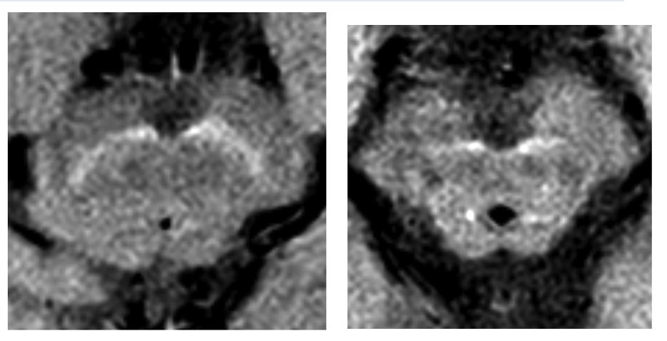
10/ The area and contrast ratio of T1 high-signal NM pigmentation is lower in patients with PD (example on the right) in comparison to healthy controls (example on the left).
1
0
1
8/ Neuromelanin (NM) imaging makes use of the fact that neurodegeneration in PD with dopaminergic cell loss causes reduced NM-pigmented neurons. NM-sensitive MRI uses high spatial-resolution T1-weighted imaging with fast spin-echo sequences at 3T.
1
0
0
7/ Nigrosomes 2-5 can be identified on high-spatial resolution MRI scans. On 3T SWI, nigrosome hyperintensity loss in PD progresses from nigrosome 1 to 4 with later stage PD.
1
0
0
6/ PD patients show loss of that hyperintensity (example below) on SWI. At 3T, this sign is highly sensitive (94.6%) and specific (94.4%). In addition to PD and Parkinson-plus syndromes, loss of nigral hyperintensity can be seen in rapid eye movement sleep behavior disorder.

1
0
0
5/ In PD, dopamine loss in the SN occurs in the nigrosomes or calbindin-poor zones within calbindin-rich neutrophils of the nigral complex. Maximal cell loss occurs in nigrosome 1, shown in red below. “Swallow tail sign” refers to the hyperintensity on SWI.

1
0
0
4/ Several imaging tools have emerged for visualization of the SN, which can detect findings characteristic of PD, monitor progression, and differentiate PD from other syndromes. 1)Nigrosome imaging .2)Neuromelanin imaging .3)Quantitative Iron Mapping.4)Diffusion Tensor Imaging.
1
0
0
3/ ANSWER: Essential Tremor. Nigrosome imaging demonstrates loss of normal hyperintensity in PD and Parkinson-plus syndromes. Drug-induced parkinsonism, dystonic tremor, essential tremor, and vascular parkinsonism will have an intact nigral hyperintensity.
1
1
1
2/QUESTION: Loss of the “Swallow-tail” sign on susceptibility weighted imaging (SWI) is LEAST likely to be seen with which of the following entities?.
1
0
0
So excited to have matched at @UMichRadiology for #Neurorad fellowship! Excited to be going back home to the Midwest and joining @cmadamanchi . #goblue

12
1
103