
Aaron Rutman, MD
@aaronrutman
Followers
4K
Following
3K
Media
311
Statuses
760
Neuroradiologist, @kpwashington, @UWRadiology, UW SOM #neuroanatomy instructor; photography, guitar, baseball, dad of 3 #neurorad
Seattle, WA
Joined June 2014
Reposting #neuroanatomy #neuroradiology tutorials for my fall semester #medicalstudents. #meded #FOAMed #FOAMrad #medtwitter #medstudents #radiology #neurorad #radres #neurology #neurosurgery
1
7
37
RT @MohitTCCNeuro: Join us for the next webinar in @thecortexclub T-bone series, on Thursday Jan 23 at 11AM Central US time, with the outst….
0
19
0
RT @thecortexclub: Register here: for this webinar on Jan 23, 11AM Central US time by none other than @amyfjuliano….
0
8
0
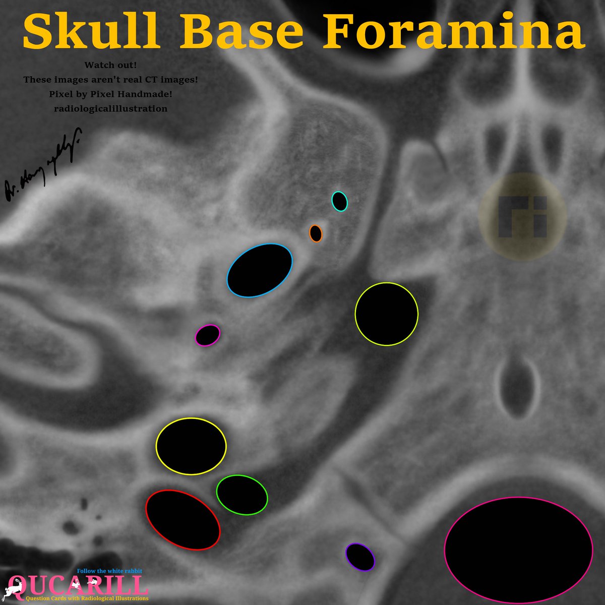
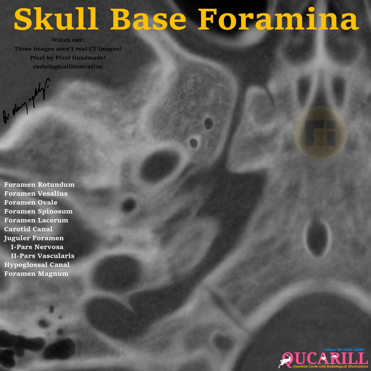
RT @drharunyildiz: Skull Base Anatomy . #radiologicalillustration ✍️.pixel by pixel handmade!


0
121
0
RT @AMoosaMD: Cool. I gave a similar talk (less MRI and more functional Neuro anatomy to our trainees in epilepsy, and neurology @Cleveland….
0
30
0
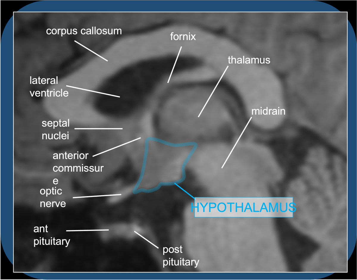
Hypothalamus! #anatomy #neuroanatomy.
HYPOTHALAMUS (HT)🧵-the control center of circadian rhythm, fatigue/wakefulness, hunger/satiety, sex drive, thirst/BP—and the command post for endocrine control via the HT-pituitary axis. #meded #neuroradiology #neuroscience #radiology #neurology #neurosurgery #neuroanatomy.1/28
0
3
5
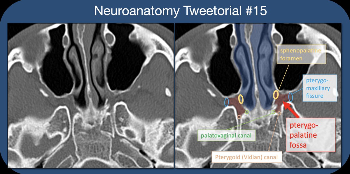
Pterygopalatine Fossa! #anatomy #neuroanatomy.
Pterygopalatine fossa🧵- inspired by ?s from med students in neuroanatomy lab & a resident w/ case of perineural tumor spread on same day! #meded #FOAMrad #medtwitter #medstudents #radiology #neurorad #HNrad #radres #neurology #ENT #temporalbone #neurosurgery #neuroanatomy.1/22
1
3
12
A case illustrating oculomotor anatomy! .
A plea to all new&old #medstudents and #residents: an imaging study is a consult with a sub-specialized physician, not a lab test. Providing appropriate indications is 🔑->get the right study done & get it read with appropriate pre-test probabilities considered! #radiology. 7/13

1
1
2
ICA branches!.
Neuroanatomy TOTD #6.a) Ant choroidal artery infarct, involving the post limb of the internal capsule.b) Sup hypophyseal artery aneurysm—medially directed from the supraclinoid ICA.#meded #FOAMed #FOAMrad #radres #neurorad #radiology #neurosurgery #neuroanatomy #neuroanatomyTOTD


1
1
2
Vidian/pterygoid canal!.
Neuroanatomy TOTD #1.1/7 The arrows point to the bilateral pterygoid canals (aka Vidian canal), seen here on a coronal CT of the skull base. Note that the canal travels in the AP direction, within the sphenoid bone, medial and inferior to the foramen rotundum.

1
1
1
Find the Central Sulcus!.
Neuroanatomy TOTD #3.1/6 The superior frontal sulcus courses in the AP direction, and terminates at the precentral sulcus posteriorly. The central sulcus is immediately posterior to the precentral, and the postcentral immediately posterior to the central. #meded #FOAMed #FOAMrad

1
1
2
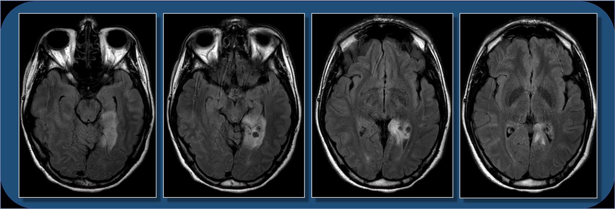
Inferior brain gyri.
Hey #radres and #MEDstudents - a #neurorad case for you today. 18yoF w/ seizure. What do you see? What is the ddx? And WHERE is the lesion? Case explanation and gyral #neuroanatomy explanation coming soon. #meded #medtwitter #FOAMed #FOAMrad #radiology #neurology #neurosurgery
1
1
2
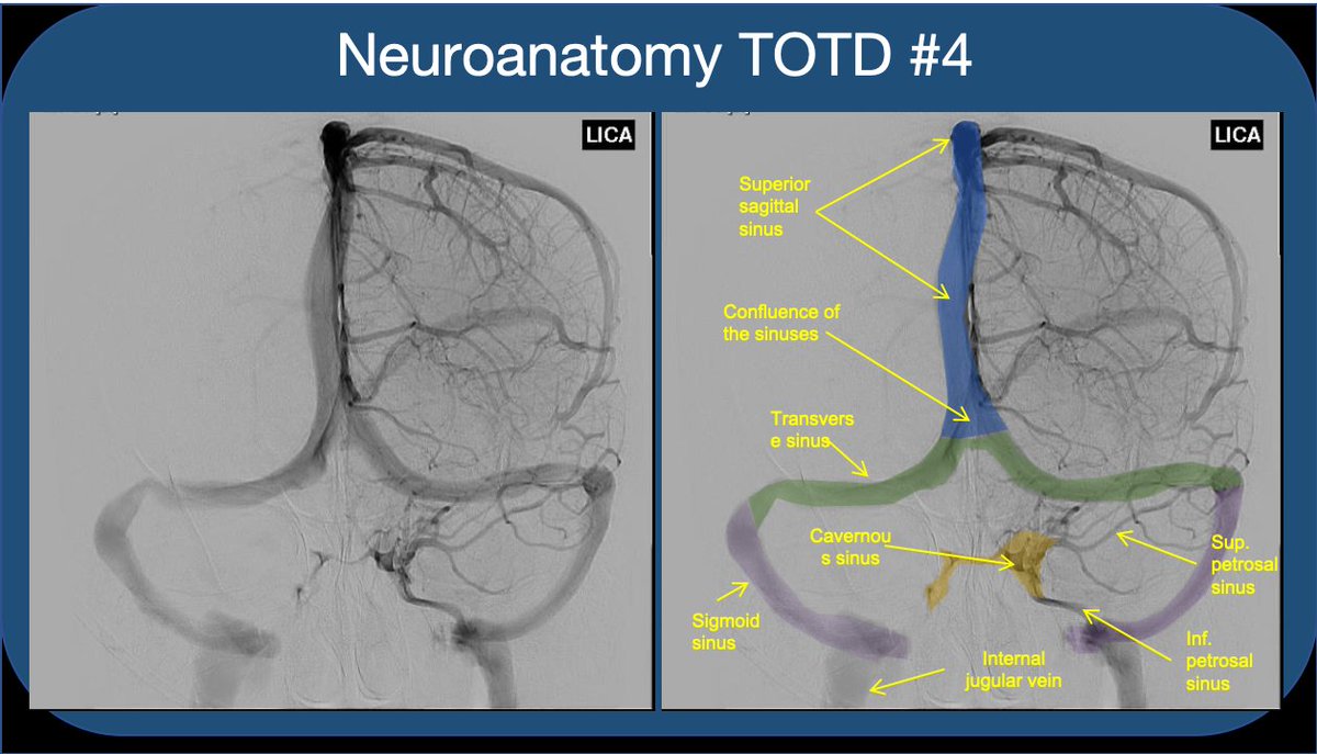
Cavernous Sinus!.
Neuroanatomy TOTD #4.1/5 Answer: The orange structure is the cavernous sinus (CS), a paired dura-lined venous cavity on either side of the sella. The sinuses are split into numerous “caves” by fibrous septae (hence the name). #neuroanatomy #neurorad #medtwitter #neuroanatomyTOTD
1
1
1
Facial Nerve (CNVII)!.
Neuroanatomy TOTD #11🧵.➡️intracanalicular facial nerve. IAC cross section➡️4 nerves; ant-sup➡️facial, ant-inf➡️cochlear (7-up/coke-down). Post nerves➡️sup&inf vestibular #medtwitter #meded #FOAMed #FOAMrad #neurorad #radiology #radres #neurology #neurosurgery #neuroanatomy.1/11

1
1
1
Basal Ganglia!.
Neuroanatomy TOTD #10🧵 .1/5 Small gray matter structure at the junction of the thalamus and midbrain is the subthalamic nucleus (STN). #meded #FOAMed #FOAMrad #medtwitter #medstudents #radiology #neurorad #radres #neurology #neurosurgery #neuroanatomy #neuroanatomyTOTD

1
1
2
Meckel's Cave (CNV)!.
Neuroanatomy TOTD #9🧵.1/6 The trigeminal n. courses anteriorly➡️prepontine cistern➡️into Meckel’s cave (green), which lies at the medial floor of middle cranial fossa at the petrous apex. #meded #FOAMed #FOAMrad #neurorad #neurology #neurosurgery #neuroanatomy #neuroanatomyTOTD

1
1
0
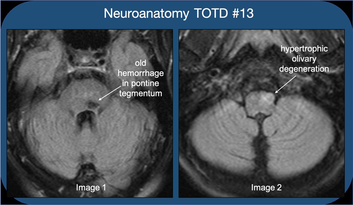
#NeuroanatomyTOTD #13🧵. Image1: hemosiderin due to old hemorrhage in the L pontine tegmentum. Image2: ipsilateral hypertrophic olivary degeneration (HOD) . 1/13. #meded #FOAMed #FOAMrad #medtwitter #medstudents #radiology #neurorad #radres #neurology #neuroanatomy #neuroscience
1
1
2
Amygdala/stria terminalis/limbic system!.
Neuroanatomy TOTD #12. The green structure is the amygdala (amygdaloid body) and the yellow structure is the stria terminalis (ST). 1/18. #meded #FOAMed #FOAMrad #medtwitter #medstudents #radiology #neurorad #radres #neurology #neurosurgery #neuroanatomy #neuroanatomyTOTD

1
1
1
Hippocampus!.
Neuroanatomy TOTD #8🧵.1/8 MRI shows the hippocampal formation; the hippocampus(HC) is a ridge of archicortex gray matter at floor of lat ventricle, in the med temp lobe #FOAMed #FOAMrad #radres #neurorad #neuroscience #medtwitter #radiology #neurology #neurosurgery #neuroanatomy

1
1
2
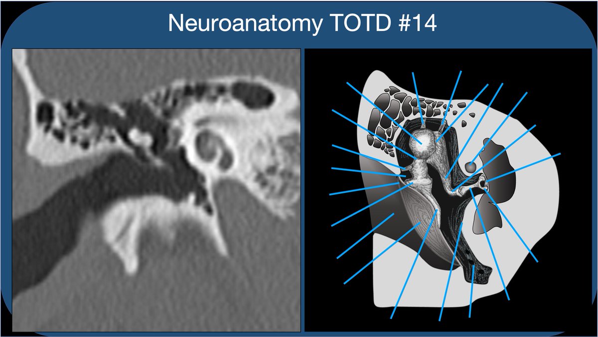
Middle Ear!.
Neuroanatomy TOTD #14🧵 .Got some requests to do one of the trickiest areas of human anatomy, the #temporalbone. So many named structures! #meded #FOAMed #FOAMrad #medtwitter #medstudents #radiology #neurorad #radres #neurosurgery #neuroanatomy #ENT #otolaryngology . 1/21
1
1
1
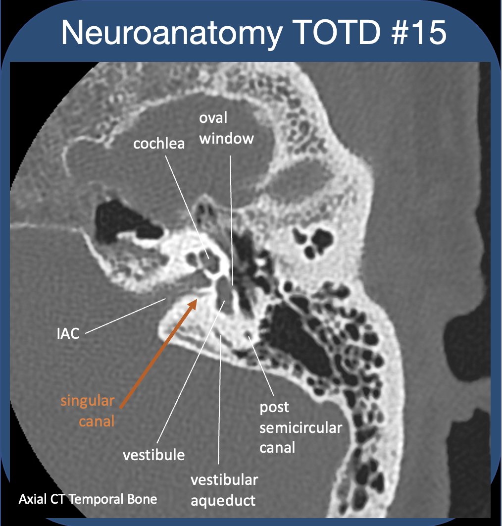
Inner Ear!.
Neuroanatomy TOTD #15🧵 .The inner ear #tweetorial--it packs a large functional punch for its small size-strap in!.#meded #FOAMed #FOAMrad #medtwitter #medstudents #radiology #neurorad #HNrad #radres #neurology #ENT #temporalbone #neurosurgery #neuroanatomy #neuroanatomyTOTD.1/24
1
0
3