
Divya Gunda
@learnneurorad
Followers
6K
Following
4K
Media
177
Statuses
567
Neuroradiologist @CooperRadRes, here to share #Neurorad cases for #radres #RadEd. Tweets my own, for #MedEd, not medical advice. Alum: @PennRadiology @OUHealth.
Joined September 2015
This is an easy one for most but still see this overlooked and it helps to see it many times. History of generalized seizures. Findings of left mesial temporal sclerosis with significant atrophy and abnormal signal in the left hippocampus. Image is not pretty but that’s reality!

1
4
24


3
8
37
The best response to my “announcements”.

@learnneurorad @drharunyildiz we all know its considered a normal anatomical variation rather than a pathological finding. no need to announce that.
0
1
1
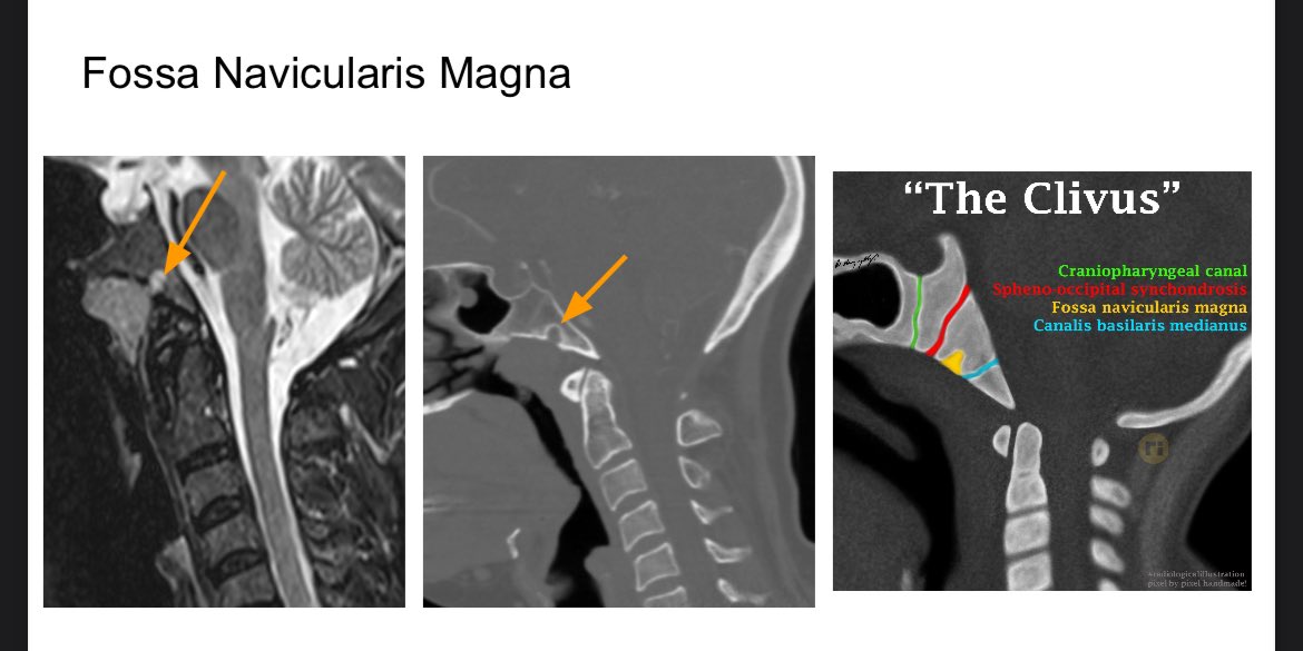
Fossa navicularis magna is a benign developmental lesion manifesting as an indentation in the anterior inferior clivus. This does not need follow up, biopsy or further imaging. @drharunyildiz illustration of different developmental lesions of the clivus. #Neurorad #radres
5
56
262
The next step in management in this patient is nothing. This is a fossa navicularis magna in the anterior inferior clivus, which is a developmental lesion related to notochord migration and does not have a propensity to transform into anything else.
0
1
9
What is the next step in management for the lesion in the clivus?.
1
0
4

The hyperdensity in the right frontal lobe is concerning for hemorrhage in the setting of trauma. However, the lack of edema should raise the possibility of a hyperdense lesion, such a cavernous malformation. Follow up mri demonstrates a popcorn lesion on T2 with adjacent DVA.

2
6
47
What is the next step in management in this trauma patient?.
2
0
11

14
18
117
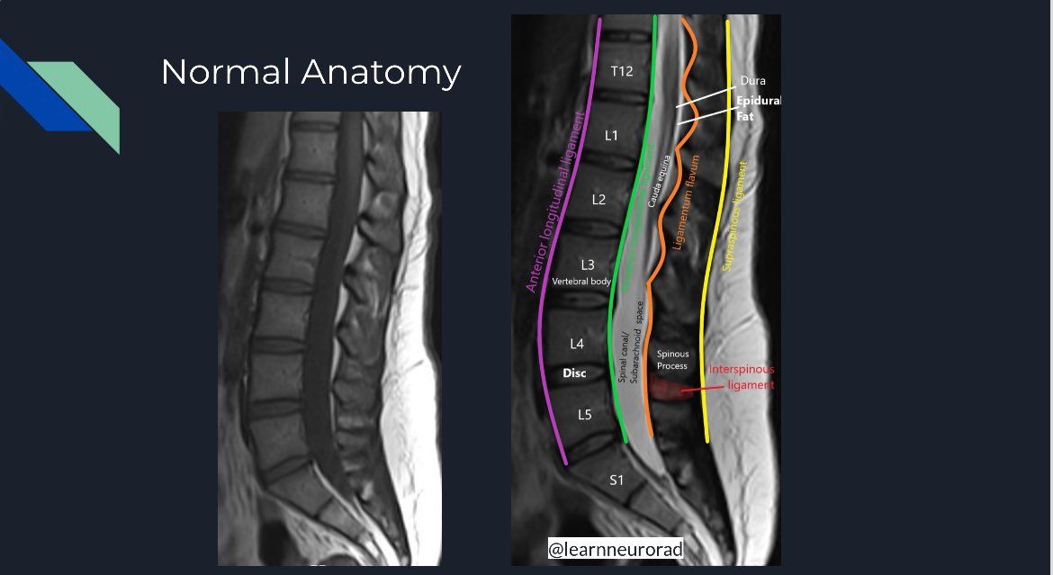
RT @learnneurorad: Normal anatomy of the mid-lumbar spine at the L3-4 intervertebral disc level in the axial plane on a T2-weighted MRI. Th….
0
67
0


MRI brachial plexus shows a distal thoracic duct draining into the IJ & subclavian vein confluence. It can often have a flared or tubular configuration, not to be mistaken for pathology such as lymphadenopathy or brachial plexus injury, which would be more posterior. #radres


1
11
59

RT @AAguilera_MD: Important take aways from Dr. Divya Gunda’s talk on the jugular foramen and foramen magnum. #ASHNR24 #HNrad #neuroradiolo….
0
13
0
RT @ASHNRSociety: Beautiful overview of some of the intricate anatomy of the jugular foramen with @learnneurorad #ASHNR24 .
0
32
0


RT @MohitAgNeurorad: Come join us tomorrow Sep 04 at 10:15am to see these 10 superstars in action at the #ASHNR24 @ASHNRSociety T4C - @thec….
0
5
0