Leica Microsystems
@LeicaMicro
Followers
16K
Following
3K
Media
2K
Statuses
7K
Leica Microsystems is a world leader in #microscopy and scientific instruments. Instagram: https://t.co/lq5PpCGu3g LinkedIn: https://t.co/bVniUzFoSf CEO: @LeicaMicro_CEO
Wetzlar, Germany
Joined February 2011
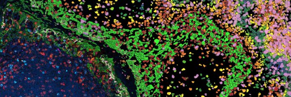
Researchers have now discovered how many #peroxisomes (an organelle which breaks down toxins and makes vital molecules) a cell needs is regulated by an enzyme called PKC. Explore the research ➡️ . This @BPoD_s was created using @LeicaMicro microscopy.

0
2
4
Leica Microsystems is proud to be part of @DanaherCorp.As part of Danaher, we accelerate the power of science and technology to improve human health and deliver innovation at the #speedoflife. Learn more:
0
0
2

Quickly capture sample details with a press of a button while keeping your eyes on the image with the Visoria B laboratory microscope. The button for image acquisition is easily accessible on the microscope stand. Simplify your documentation w/ Visoria B:
0
1
4
⌛️ Want to improve your inspection efficiency? Discover the top challenges during #VisualInspection using a microscope (and how to overcome them) in our article: “Top Challenges for Visual Inspection.” 👉️ . #QualityControl #InspectionSolutions

0
1
1
Step into the world of discovery! 🔬 Hear from @MBLScience summer courses participants as they share their research, cool techniques they’ve mastered & advice for future learners. Full interview 👉️ Thank you Ryo, Vinaya & Bharat!.#ScienceStory #MBL2025
0
0
6
RT @NeuroAlc: #SciencePhoto_IN.“Echoes of a neural storm”.Author: Carlos Avilés Granados.Lab: Dr. Javier Sáez Valero."Post-mitotic neurons….
0
1
0
RT @NeuroAlc: #SciencePhoto_IN.“Retina and retinite: a love story”.Author: Isabel Pérez Ferrer @isaabeel_24.Lab: Dr. Eloísa Herrera @LabEHe….
0
3
0
🔬 Free webinar Sept. 9! Discover the future of label-free chemical imaging w/ #STELLARIS CRS. See order-of-magnitude faster chemical imaging powered by advanced laser technology - integrating #SRS, #SHG, two-photon fluorescence & more. Sign up:
0
0
1
Do you evaluate the optical properties of #anisotropic materials? The Visoria P polarization microscope offers three modules for #conoscopy, such as Bertrand lens cube, Bertrand lens module (A/B module), and Advanced conoscopy module. Learn more:
0
1
2
📢 @LeicaMicro is excited to announce a new partnership w/ @fishersci to expand access to our standard compound & stereo microscopes across EMEA! Empowering scientists and researchers where speed, convenience & reach matter most. More info:
1
0
4
RT @NeuroAlc: #SciencePhoto_IN.“Floral neurogenesis”.Author: Carlos Avilés Granados.Lab: Dr. Javier Sáez Valero."Neural progenitors (nuclei….
0
1
0
RT @NeuroAlc: #SciencePhoto_IN.“Flying Memory”.Author: Mirjam Cangonja.Lab: @BarcoLab."Viral expression selectively labels hippocampal exci….
0
2
0
Look beyond what you've imagined. Discover #MyVeo, the all-in-one surgical visualization headset for #ophthalmology! 🔗 .✅ Unify essential real-time clinical data right in front of your eyes.✅ Stay focused, work comfortably & enhance team collaboration

0
0
3
Join our Lunchtime Lectures at MC 2025! Don’t miss these sessions:.🔬 UC Enuity Cryo Workflows.🔬 Battery Workflows.Reserve your seat now to secure your lunchbox! . #CryoEM #BatteryResearch #Ultramicrotomy #MC2025

0
0
1
Adapt the Visoria M materials microscope to the way you work! Its ergonomic accessories let you customize the microscope’s height, viewing angle, and eyepiece position, enhancing comfort and flexibility for long hours at your workstation. 🔬 More:
0
0
1
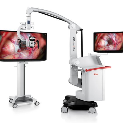
The ARveo 8x is designed to protect delicate tissue w/ Advanced Illumination:.🔦 BrightCare Plus auto-optimizes light intensity based on working distance.🔦 AutoIris adjusts the diaphragm to illuminate only the visible surgical area. 👉️ More:
leica-microsystems.com
The ARveo 8x hybrid surgical microscope provides you with our best optical visualization for neuro, spine, and plastic reconstructive surgery. Digital capabilities and 3D-visualization technology....
0
0
3
RT @NeuroAlc: #SciencePhoto_IN.“The Lung Bird: A Tale of Colonization”.Author: Angelita Costantino.Lab: @NietoAngela."Metastatic breast can….
0
1
0
"What really makes the difference for me? It’s the people behind the products." – Philipp Becker, Sr. Manager Assembly Ops.Curious about a career at #LeicaMicrosystems?.Check out our career site and be a part of our innovative journey! .👉. #IamLeica

1
0
0
Watch our on-demand webinar to learn how you can streamline your visual inspection workflow to optimize efficiency ➡️.🔬 Learn from Leica expert, Michael Doppler, with over 30 years of microscopy experience. #Electronics #PCB #Semiconductor

0
0
0
Heading to #MC2025 in Karlsruhe? Visit @LeicaMicro at Booth 5 to see UC Enuity in action – our latest ultramicrotome for EM sample prep. 🔹 Live demos.🔹 Expert talks. Register now ➡️ .🗓️ August 31 - September 4. #ElectronMicroscopy #SamplePreparation

0
0
2