
The Neuroradiologist
@theneuroradguy
Followers
28K
Following
519
Media
237
Statuses
460
A (relatively) young neuroradiologist, fascinated by the brain and brain imaging. Here to share my passion :). #radres #RadED #FOAMrad #Neurorad #radiology
Joined December 2021


10) Thank you for reading, and if you want to know more, I made a video about it on youtube:

0
1
22
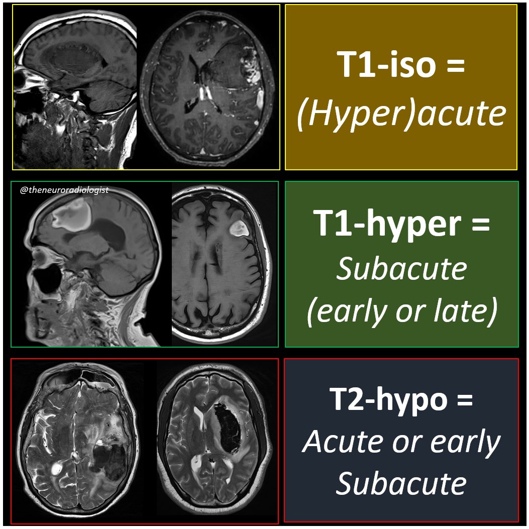
9) I know there are mnemonics, but I hate mnemonics, because I need mnemonomics ot memorize those mnemonics. This approach works better for me.
1
2
32
8) Now if you just remembrer these three rules, you can deduce all the rest and you can guess the age of any cerebral hematoma on MRI.

1
3
15
7) Rule 3: if a hematoma is hypointense on T2-weighted images, it's acute or early subacute.

1
3
13
6) Rule 2: if a hematoma is hyperintense on T1-weighted images, it's subacute (early or late).

1
3
16
Rule 1: if a hematoma is iso-intense on T1-weighted images, its (hyper-)acute.

1
3
16
But it's not. First of all, to guess the age of a cerebral hematoma, you just need T1- and T2-weighted sequences (and forget about the chronic phase, that's just some hemosiderin stained tissue loss, no longer a true hematoma).

1
4
17
This is a simplified overview of the evolution of cerebral hematomas on T1-, T2-, SWI- and diffusion-images. Looks complicated no?

2
7
44
2) Now first of all, keep reading even if the next tweet might scare you a bit.

2
0
12


Not an "official" sign (but when do radiological signs become official?) but this likekness was brought under the world's attention in an article on neuroradiological imaging signs by Inna Page and @frankgaillard (.

0
0
11
I think trident sign is the best known sign and probably the oldes so didn't bother looking up the first publication of it. In somewhater older literature also the boring name "inverted omega sign" is sometimes used.

1
1
10
This sign was first described in 2009 by Judith Wagner and colleagues in Clinical Neuroradiology .

1
0
4
Hard to find the first description of the owl sign, or "owl eyes appearance". First mention I found was on an EPOS poster from 2011 on animals in the brain:

1
0
7
As far as I know this "sign" was first published in 2009 by A González-Aguilar, is not widely known or used, but I found some mentions of it in review articles.
1
0
6

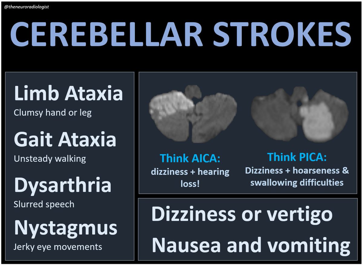
Cerebellar strokes often present with non specific symptoms like dizziness, vertigo, nausea and vomiting. Diagnosis often less straight forward than in anterior circulation strokes.
0
14
89
The most dramatic clinical presentation of brainstem stroke is no doubt the locked-in syndrome, seen in complete midbasilar artery strokes. in incomplete midbasilar strokes presentation can be more variable.

1
14
62