
Bement Lab
@BementLab
Followers
712
Following
46
Media
44
Statuses
88
Transient contractile arrays. Part of the Center for Quantitative Cell Imaging (@UWCQCI) at UW-Madison. Tweets mostly by @ZacSwider and @AniMichaud1.
Madison, Wisconsin
Joined March 2019
Join @AniMichaud1 on a tour of the Bement lab @UWMadison and see what we're all about! #CellBiology
https://t.co/6a1EnNAdJY
1
4
27

Any #microscopy friends have any suggestions for a water stage cooler/pump? I'm specifically interested in something to slowly flow water through a light sheet imaging chamber so it will need a very slow and consistent flow rate. Maybe @ZakSwartz ?
2
1
0
How does RGA get recruited to cortical waves and what exactly is the nature of its relationship with F-actin? We aren't sure, but we plan to use immature frog oocytes and modeling to address these questions!
1
2
28
Modeling and experiments showed that Ect2 and RGA-3/4 are major drivers of this circuit, and just altering the relative levels of these two players produced a variety of cortical behaviors in an otherwise quiescent oocyte cortex!
1
3
3
In summary we found that frog and starfish excitability is comprised of a circuit involving Ect2, Rho, F-actin & RGA-3/4, with RGA-3/4 regulating negative feedback along with F-actin in a manner still mostly unknown...
1
0
2
We concluded that activated starfish oocytes and embryos and immature frog oocytes represent distinct phases on this excitability spectrum. Immature frog oocytes occupy the lower end while activated starfish oocytes and embryos occupy the higher end.
2
2
1
As the model ramped up the amount of RGA and entered the oscillatory domain, we saw an increase in amplitude and morphological features, and then a gradual decrease. In vivo experiments in oocytes closely paralleled these findings.
1
3
8
Modeling and experiments revealed two uniform states with no waves, separated by an oscillatory domain that supported a variety of cortical wave behavior. Bounding the oscillatory domain were regions of wave instability, characterized by pulsatile and spiral turbulence.
1
1
3
We were perplexed as to why RGA had a negative effect on Rho dynamics in activated starfish oocytes and embryos but was required for high-amplitude waves in the immature frog oocyte model, so we turned to theoretical modeling in combination with some in vivo experiments...
1
1
3
We used the immature frog oocyte system to look at some potential players in the excitability model: Anillin, myosin-2 and Dias 1-3. We showed the oocyte is a useful tool for studying these proteins further in the context of excitability.
1
1
9
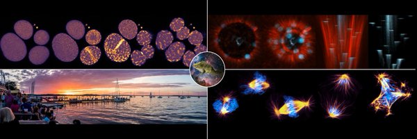
Essentially, we took the wave dynamics seen in the embryonic furrow and induced them over the quasi-two-dimensional space of the oocyte cortex. This not only looks cool, but also allows us to collect high spatiotemporal data without complications present in embryos.
1
8
19
Light sheet imaging with @henryhetired and @spim revealed that these patterns were wide-spread and well-developed at merely 5hrs post-mRNA injection. This behavior was not seen with GAP-dead RGA or another cytokinetic GAP (p190RhoGAP).
2
35
102
To do this, we induced excitability in a cell type that does not naturally display it: the immature frog oocyte. We found that while Ect2 or RGA alone did not induce sustained excitability, co-expression of both led to high-amplitude, psychedelic waves that covered the cortex.
2
1
15
These results (and others) led us to conclude that Rho activation is mediated by Ect2, while Rho inhibition depends on RGA-3/4. Our theoretical modeling indicated that these two proteins were the backbone of our cortical excitability circuit, so we decided to put this to the test
1
1
2
A low-dose of Latrunculin B that altered (but did not abolish) F-actin waves caused a corresponding change in RGA-3/4 dynamics. RGA waves became sharper and thinner, while Rho waves became thicker and brighter.
1
0
6
Over-expressing RGA-3/4 in waving starfish cells lead to a decrease in Rho wave amplitude, and width, suggesting a role in negative feedback.
1
2
4
In starfish and frog, we found that RGA localized to the contractile ring and formed dynamic cortical waves that trailed Rho activation waves, overlapping extensively with F-actin.
1
3
18
RGA-3/4 has previously been shown to negatively regulate Rho activity during both cytokinesis and the pulsed contractions of early C. elegans embryos (Michaux et al. 2018), so we were curious if it played a role in cytokinetic cortical excitability.
1
0
1
Rho activity propagates via an Ect2-dependent, positive feedback loop, and is snuffed out by a delayed, F-actin dependent negative feedback loop. In this paper, we found that the Rho GAP RGA-3/4 plays an important role in regulating the negative feedback loop.
1
0
2
This is clearly evident in frog and starfish cells, where dynamic waves of Rho activity and F-actin polymerization traverse the cortex and get focused at the cell equator by the anaphase spindle (Bement et al. 2015).
1
1
8
Rho GTPases are key regulators of cortical excitability, and drive downstream cortical remodeling via interactions with GEFs, GAPs and effector proteins. In contrast to their classic branding as static "on" and "off" switches, GTPases are commonly deployed in dynamic patterns.
1
0
1