
Jianghao Liu
@jianghao_liu_
Followers
245
Following
261
Media
50
Statuses
217
Cognitive neuroscience of visual mental imagery, aphantasia and consciousness.
Paris, France
Joined January 2019
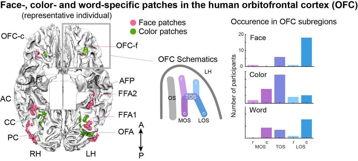
(1/9) Thrilled to share our new preprint🧠Using 7T fMRI, we report the presence of face-, color- and word-specific patches in the human #orbitofrontal cortex (OFC), which form continuous functional gradients with VOTC patches.
1
8
26
Does aphantasia have unconscious imagery? .Thrilled to annonce our new paper out on @CurrentBiology .
We provided three criteria following the current definition of unconscious imagery by @BenceNanay, and examined the emerging neuroimaging evidence on aphantasia.

0
0
5
Differences: 1. although aphantasics encoded stimulus content during imagery, these representations differed from ones elicited by perception. 2.aphantasics exhibited reduced connectivity between anterior visual areas and the OFC patches (exert top-down modulation in visualisers)

0
0
0
A follow-up 7T fMRI study on face- and colour-processing systems in aphantasia revealed normal ventral temporal activity. see 🧵.
(1/5)Category-specific visual processing in #aphantasia? .Using 7T fMRI, we systematically examined face and color patches and found that aphantasics have normal activity in the VOTC visual cortex, but deficits in (top-down) OFC activity.

1
0
1
Thus, we propose that the neural basis of aphantasia involves a functional disconnection between high-level visual regions in the ventral temporal cortex and left prefrontal areas. And the precise nature of hemispheric interactions in aphantasia warrants further investigation.
1
0
0
Aphantasia has a higher activation of the #ventralAttentionNetwork in both imagery and perception (connected with the left imagery network), this aberrant activation might heighten susceptibility to external interference, potentially either destabilizing or suppressing imagery.

1
0
0
Critically, the FIN was functionally disconnected with the left anterior PFC across domains, and with left FEF for some domains. This is highly potentially related to visual #awareness of internally generated experience in aphantasia (see above TICS letter).

1
0
0
In both imagery and perception, both groups activated a specific left fusiform area, termed Fusiform Imagery Node (FIN), across domains. While the representation of the same stimuli in imagery and perception was correlated in visualisers, this overlap was absent in aphantasia.

1
0
0
During imagery attemps, in each domain, they have corresponding domain-specific areas activated in the high-level visual cortex, such as FFA for faces, vWFA for words, color-biased areas, PPA for map.

1
0
0
What neural mechanisms create this disparity between subjective experience and objective performance in aphantasia?.
1
0
0
Congenital aphantasia struggles to visualize objects despite being able to describe their visual appearance. In both imagery and perceptual tasks, their RTs were slower and they had lower confidence in their responses on perceptual tasks.

1
0
0
🚨Out in @TrendsCognSci, ."Aphantasia as a functional disconnection"!.A disconnection between the Fusiform Imagery Node (FIN) & the left PFC may explain retained memory for objects, despite lacking subjective imagery in aphantasics. left PFC, awareness, abnormal attention. 🧵

1
4
21
Impressive study! The fusiform imagery mode (FIN) is disconnected in acquired aphantasia!.

Can brain injury cause the loss of visual imagination? . We studied #aphantasia due to brain injury. Lesions were in many different regions but 100% were connected to fusiform imagery node - a region active during visual mental imagery @Brain_Circuits .

0
1
5
RT @TomerUllman: Out now in TiCS, a thing i've been thinking about a lot: . "Physics vs. graphics as an organizing dichotomy in cognition"….
0
17
0
RT @GretaTuckute: What are the organizing dimensions of language processing?. We show that voxel responses are organized along 2 main axes:….
0
40
0
(5/5)Thus, during imagery attempts, aphantasics could activate the visual cortex to represent stimulus-specific content, albeit different to that employed during visual perception. The OFC in aphantasia may be related to deficits in the top-down modulation of imagery experience.
1
1
1
(4/5) During imagery maintenance period, aphantasics showed reduced activity in the #OFC, but not in any areas of the visual cortex. (Could this be related to a "only flash of imagery" experienced by some aphantasics?🤔

1
0
1
(3/5) However, there are #three group differences. 1. although aphantasics encoded stimulus content during imagery, these representations differed from those elicited by perception. 2. aphantasics exhibited reduced connectivity between the OFC and anterior visual areas.

1
0
1
(2/5) Interesting, during imagery attempts, in the visual cortex, aphantasic participants i) showed activation in relevant face-, and color-specific patches (instead of being silent); ii) encoded visual content (e.g. visual color appearance).😲.
1
0
1
(1/5)Category-specific visual processing in #aphantasia? .Using 7T fMRI, we systematically examined face and color patches and found that aphantasics have normal activity in the VOTC visual cortex, but deficits in (top-down) OFC activity.
1
0
2