
@Hemepathguy
@hemepathguy
Followers
5K
Following
3K
Media
427
Statuses
1K
The real, former @DennisOMalleyMD. Opinions are NOW my own. Hematopathologist, #lymphoma, #heme, #spleen, guitar
Joined May 2020
RT @PaulYoungMD1: You are invited to join our MDACC Lymphoma Unknown Conference with Dr Medeiros on Friday, June 20th at 4:00pm CST. Slid….
0
5
0

RT @PaulYoungMD1: Please join our Lymphoma Unknown Conference with Dr Medeiros tomorrow 04/18/25 4pm CST. Link to preview slides:. https://….
0
5
0
RT @PaulYoungMD1: Please join us for a high yield lecture on splenic pathology "Overview of splenic pathology" by Dr Dennis O'Malley @hemep….

mdacc.zoom.us
Zoom is the leader in modern enterprise cloud communications.
0
11
0
O'Malley DP, Lee JP, Bellizzi AM. Expression of LEF1 in mantle cell lymphoma. Ann Diagn Pathol. 2017 Feb;26:57-59. doi: 10.1016/j.anndiagpath.2016.09.016. Epub 2016 Nov 22. PMID: 28038713.
1
2
13


Did you say mantle cell lymphoma? 10% of MCL will express LEF1 (this stain is cyclin D1)

1
2
12


There is a portion of nerve and ganglion cells! Stains are calretinin (Left) and S100 (right)


0
0
17


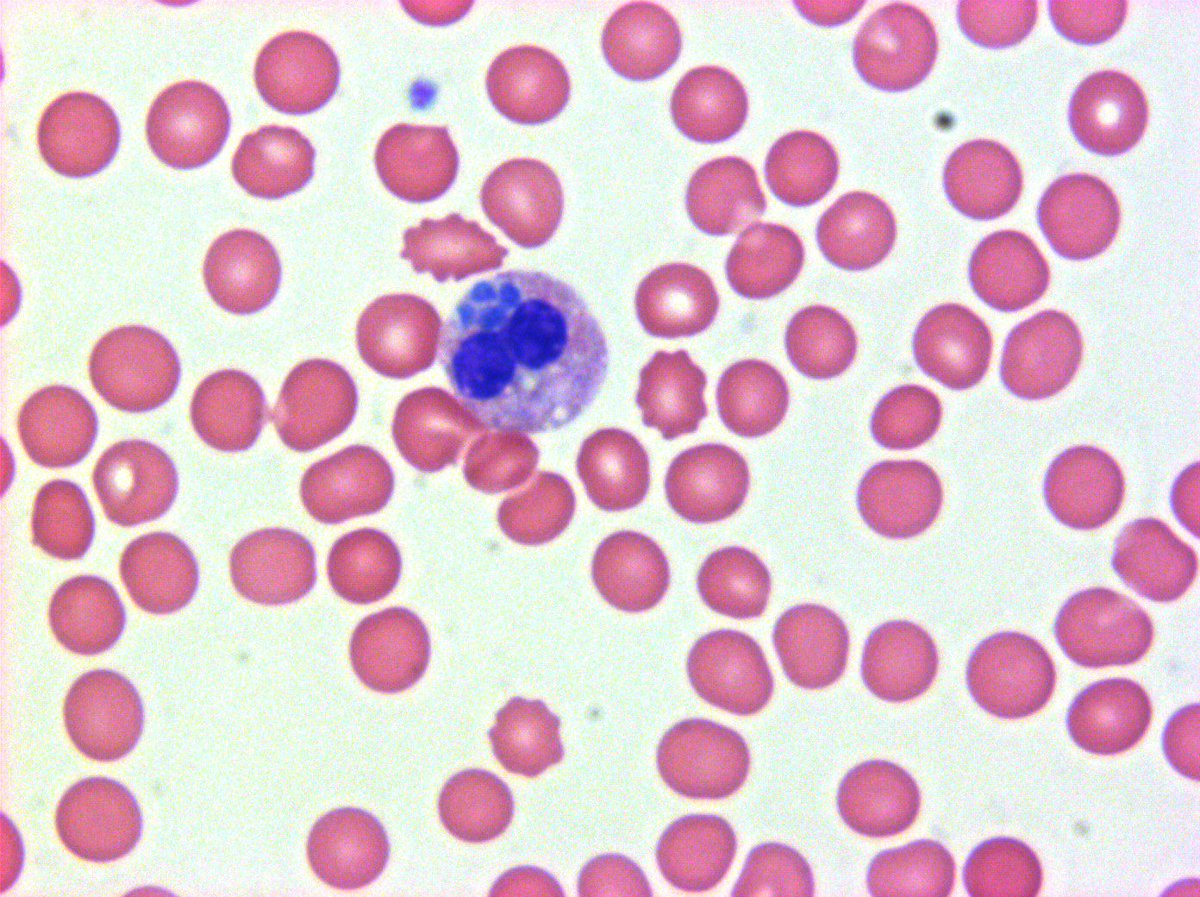
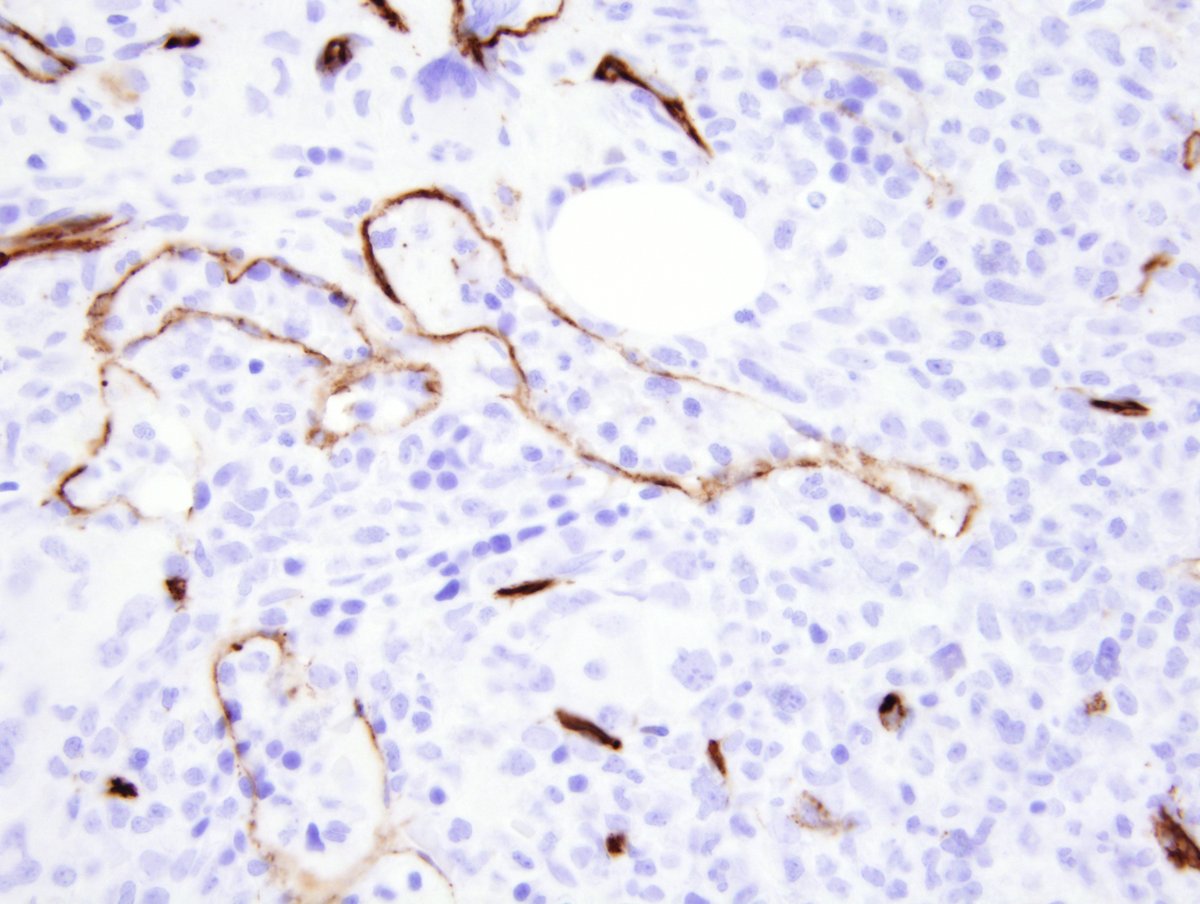
So those are megakaryocytes. The stain is CD42b. I suspect these are benign, but to be safe recommended that the patient be check for a marrow disorder, such as an MPN.
0
1
16
#hemepath 75 yo female. Axillary LN biopsy. What are those big cells? What IHC stain is shown?


3
13
48
#hemepath Splenic diffuse red pulp small B cell lymphoma. Stain is CD103 - which WHO says is "rare" but I see it occasionally


1
9
46
#hemepath Bravo to the pathologist for sampling this! Splenic artery embolization for lymphoma. This is embolization material in splenic artery!

2
11
55
RT @hemepathguy: #hemepath hepatosplenic T cell lymphoma in bone marrow. IHC are CD3 and TCR delta. Note sinusoidal involvement https://t.c….
0
21
0
RT @hemepathguy: #hemepath IHC - CD71 in marrow. Strong staining are erythroids. Weak is AML. CD71 is transferrin receptor. All proliferati….
0
17
0
RT @hemepathguy: #hemepath Somewhat historical image. This is a TRAP cytochemical stain in hairy cell leukemia

0
7
0
RT @hemepathguy: #hemepath a handful of "flame cells" from myeloma. These are most often IgA positive.


0
22
0
RT @hemepathguy: #hemepath examples of Dutcher bodies in plasma cell myeloma. They are composed of immunoglobulin that sits on top of nucle….
0
24
0