
Toby Schmitt, MD
@TobyASchmitt
Followers
1K
Following
685
Media
103
Statuses
224
Dermpath fellow | Developer of https://t.co/mXLTq4CIbQ, https://t.co/S3gFkr0ifl
Vancouver, Canada
Joined October 2022
Couldn't be more excited to announce the launch of https://t.co/iJqFUSnnfj, the virtual slide site I wish had existed during my pathology residency. You can: - Order special stains to work up cases - See annotated histologic features - Prepare for exams with board-style questions
21
103
390
A large necrotizing granuloma in the lung showing histiocytes lining central necrosis with "ghost outlines" of dead cells. * Bug stains on FFPE tissue aren't 100% sensitive (AFB and GMS were negative in this case but sputum culture showed TB) WSI: https://t.co/h3ENwciNrQ
0
4
24
A case of extranodal NK/T cell lymphoma in the uterus. - Angiocentric and angiodestructive pattern - NK cell lineage more common than T cell lineage - Positive for EBER-ISH Whole slide: https://t.co/7efUiNOhpG
1
16
58
Here's a rare indolent skin and soft tissue lesion, "ALK-rearranged myxoid spindle cell neoplasm". It's composed of whorls of cords of bland spindle cells in a myxoid background. Positive for S100 and CD34, an unusual combination. See the whole slide: https://t.co/WvUpoE0YD7
1
25
73
A section from a paraganglion showing chromaffin cells with granular basophilic cytoplasm. Unlike paraganglioma, nests don't form a discrete mass but are dispersed among background structures. Whole slide: https://t.co/RB6YWPrkfp
0
5
38
A classic gastrointestinal stromal tumor (GIST) with wavy nuclei and a background hyalinized stroma. Slide link: https://t.co/tsW8VNjpdN
0
8
43
A color negative of an H&E pathology slide (spindle cell lipoma in this case)
1
5
18
Intro to dermpath: For those learning about skin pathology, I made a collection of labelled digital slides that covers normal histology as well as the most common diagnoses you'll see day-to-day. Check it out: https://t.co/ofkWrQfrCI
2
31
147
A mucocele-like lesion of the breast (extravasated acellular mucin): -> Considered benign but some cases are associated with ADH (e.g., this case) as well as in situ or invasive carcinoma -> Recommend complete excision for partially-sampled lesions https://t.co/UayqkLEHRr
0
7
36
Lymphomatoid granulomatosis: - Small T cells with occasional large, EBV+ B cells (arrow) - Angiocentric and angiodestructive Labelled whole slide image: https://t.co/5r2UGKEuLz
0
12
61
A case of autoimmune gastritis with an "antralized" gastric body, intestinal metaplasia, and a lymphoplasmacytic infiltrate in the lamina propria. * H. pylori infection can show similar findings, so make sure an H. pylori stain is negative. 🔬WSI: https://t.co/5GXSWOHnNI
2
12
64
Smooth muscle differentiation in a low grade endometrial stromal sarcoma. See the whole slide and more info: https://t.co/HwzIv2yV2p
1
10
35
A case of a rare nasopharyngeal tumor: This slide and more were just added to https://t.co/iJqFUSnnfj, bringing us to > 1300 cases total
3
15
83
Pelvic mass, 50F. No clear attachment to any organ on imaging
2
4
32
Papillary renal neoplasm with reverse polarity: - Papillae lined by bland cuboidal cells with nuclei oriented AWAY from the basement membrane - GATA3+ See the whole slide and more info: https://t.co/3rAlL7a2Yu
0
12
46
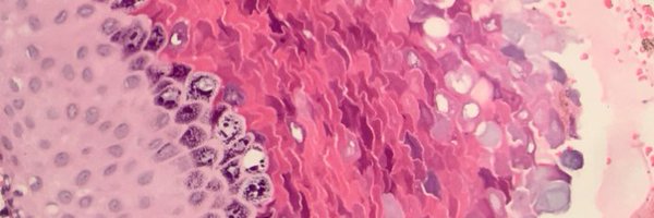
Complete hydatidiform mole: - Single population of large, hydropic villi with irregular "scalloped" borders - Trophoblast proliferation, often in a circumferential pattern around villi See the whole slide and more info: https://t.co/YAOy2uJFM4
2
20
64