
TELMA LEMOS
@TELMALEMOS10
Followers
742
Following
4K
Media
224
Statuses
2K
professor and researcher in Clinical Biochemistry post doctorate at the university of chicago - E.U.A #urinalysis#urinarysediment
Joined June 2020
RT @CarlosMartnezF7: Cytoplasmic vacuolization. Morphological change indicating cell degeneration. The origin of vacuoles is dilated organe….
0
6
0
RT @jrseltzer: Mixed cellular cast with tubular epithelial cells and WBCs - brightfield with SM stain - from patient with IgAV - #UrinarySe….
0
6
0
RT @VelezNephHepato: #UrinarySediment from pt w/newly diagnosed lymphoma and AKI. Findings suggest paraneoplastic crescentic glomerulonephr….
0
11
0
RT @edgarvlermamd: On delaying CKD progression: SGLT2 inhibitors (SGLT2i) from @goKDIGO.#NephJC #Nephpearls. #VisualGraphics by @Glomkeeper….
0
43
0

0
9
49
RT @jrseltzer: Another great #KidneyCon ! Thanks to @VelezNephHepato and @SwethaKanduriMD for running the #UrineMicroscopy workshop with….
0
6
0
RT @jrseltzer: RBC cast (also contains a couple WBCs) - brightfield with SM stain - converted to grayscale - #UrinarySediment #UrineMicrosc….
0
7
0
RT @jrseltzer: WBC cast - brightfield with SM stain - converted to grayscale - #UrinarySediment #UrineMicroscopy
0
5
0
RT @jrseltzer: Mixed cellular cast (RBC's, WBC's, and tubular epithelial cells) with a fibrin matrix - brightfield with SM stain - converte….
0
6
0
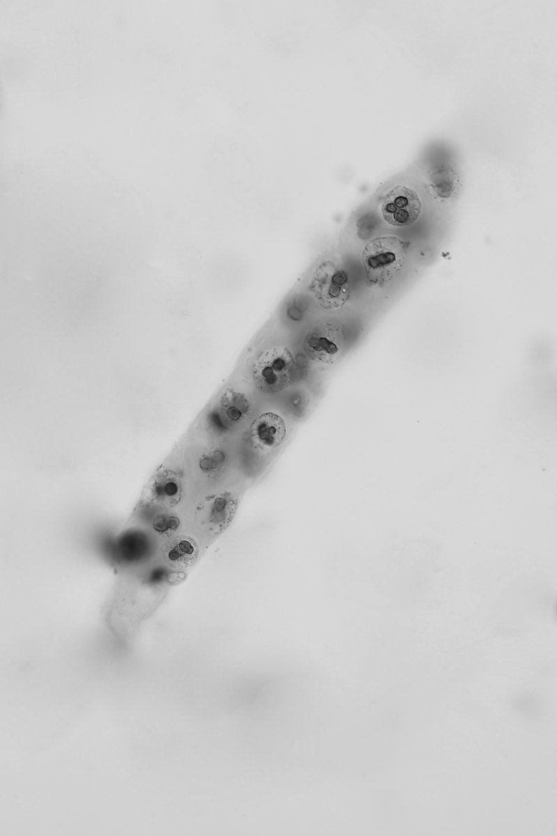
RT @jrseltzer: Renal tubular epithelial cell cast - brightfield with SM stain - converted to grayscale - #UrinarySediment #UrineMicroscopy….
0
24
0
RT @VelezNephHepato: Cellular casts, unstained r difficult to characterize it. SM stain allows better visualization but not always able to….
0
9
0
RT @jrseltzer: Mixed cellular cast, mostly RBC's, with at least one WBC - brightfield with SM stain - converted to grayscale - #UrineMicros….
0
5
0
RT @jrseltzer: Mixed cellular cast, predominately RBC's - brightfield with SM stain - converted to grayscale - #UrineMicroscopy #UrinarySe….
0
6
0
RT @QFB_Daniel: Cilindros bacterianos teñidos con Sternheimer-Malbin. Patognomonicos de pielonefritis.
0
9
0
RT @VelezNephHepato: Demonstration that nucleated cells in casts from a hyperbilirubinuric specimen are RTEC. PB stain shows that cells do….
0
7
0
RT @jrseltzer: #UrineMicroscopy samples from 2 cases today. One has bili 38 with AKI and the other a vasculitis rash, abd pain and AKI - st….
0
8
0

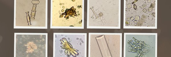
RT @CarlosMartnezF7: Different types of calcium carbonate crystals. #urinarysediment #urinalysis #urinemicroscopy

0
11
0
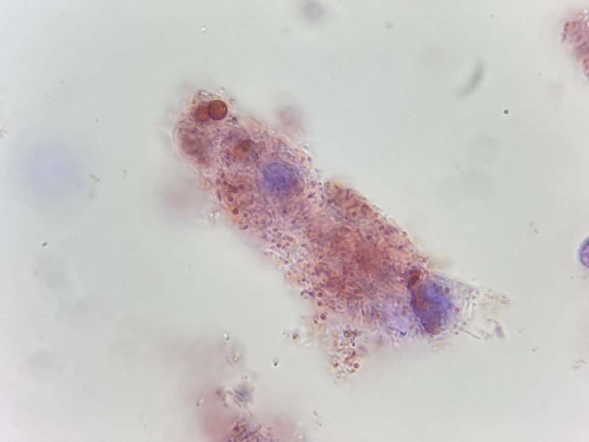
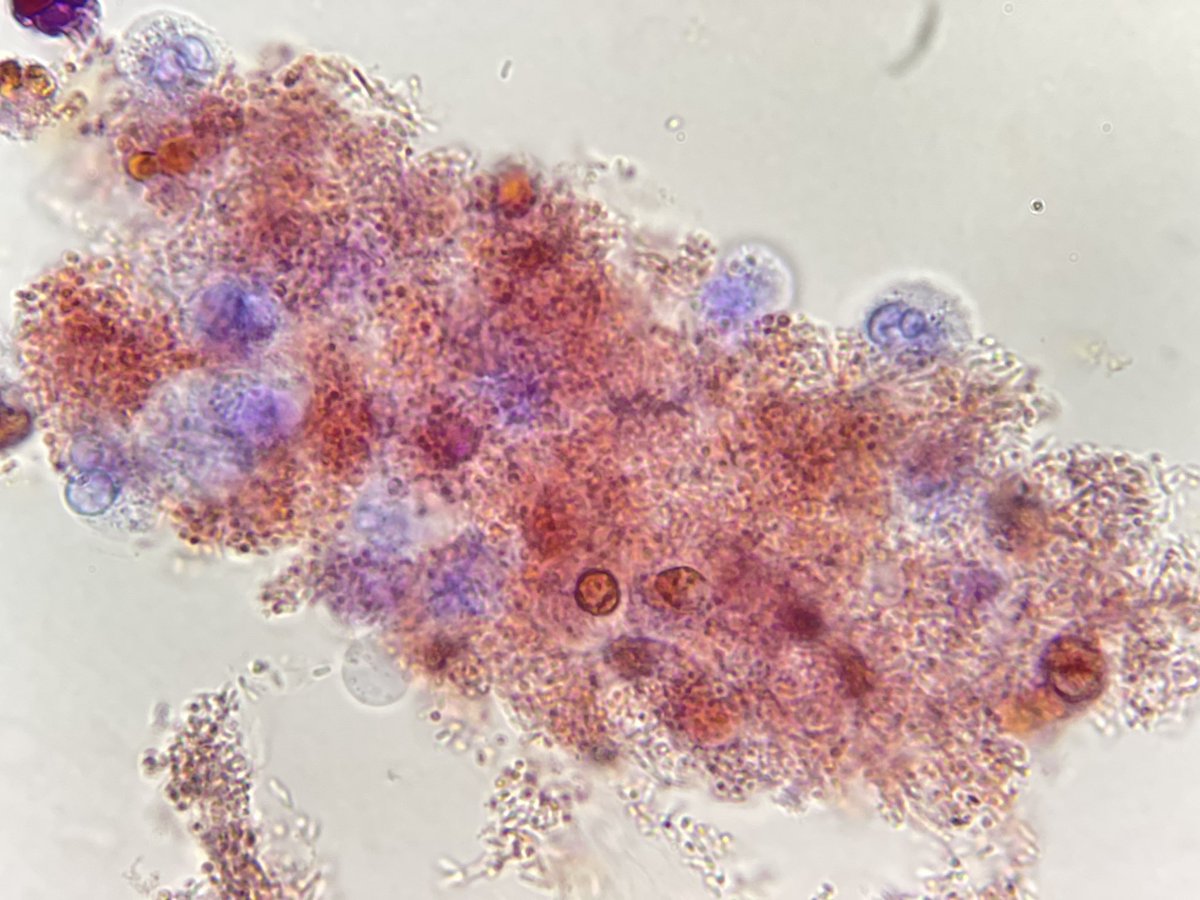
RT @CarlosMartnezF7: When a woman has vaginitis (Candida or Trichomonas) and the discharge contaminates the urine. We can observe alteratio….
0
12
0


