
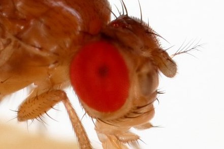
Flies are highly visual animals. That’s obvious to anyone who has ever tried to swat one. So it should be no surprise that eyes are so prominent on the fly head.
4
12
83
Replies
And that the fly visual system makes up most of the fly brain by neuron count.

1
0
3
Three months ago, the
@FlyWireNews
Consortium released the wiring diagram of a fly brain. Neuroscience finally entered the connectomic era.

We are releasing a whole-brain connectome of the fruit fly, including ~130k annotated neurons and tens of millions of typed synapses!
Explore the connectome:
Reconstruction paper:
Annotation paper:
1/6
143
1K
6K
1
0
5
The first neuronal wiring diagram for a whole brain contains—as a corollary—the first wiring diagram for an entire visual system. This wiring diagram is too complex to comprehend or even visualize, because it contains 37,000 neurons.
1
0
2
So we created a “parts list” of the 200+ types of visual neuron intrinsic to the optic lobe. Check it out at
1
0
2
I love how neurons of the Dm4 type tile the optic lobe like a kids’ playroom mat. Click for 3D interactive version.

1
3
17
Now the wiring diagram only has to describe how the 200+ types are connected, not how every single cell is connected. The entire visual system fits on a single page or in the palm of your hand.

1
0
4
Today visual neuroscience enters the connectomic era.
See preprint by Matsliah, Yu, and others from the FlyWire Consortium

1
8
32
Bonus for the deep learning aficionados: Each type of neuron is analogous to a feature map in a convolutional net. More on that coming soon...
1
0
10
ICYMI a revision of our fly optic lobe paper dropped on Monday. It’s OK, I know you were frantically finishing your 1040.

1
2
4
Just to remind you, the first version posted six months ago. It described 200+ neuronal cell types intrinsic to the fly optic lobe, and was the first “parts list” for an entire visual system.
1
0
0
We summarized the rules of connectivity between cell types in a matrix. This is the corrected version.

1
0
0
Some people told me that this matrix is pretty. But no one noticed that it was erroneously transposed in the first version. Oops!
1
0
0
Back then we claimed that the matrix was important. Six months later, we now know how to use the matrix to aid global understanding of the functional architecture of the fly visual system.
1
0
0
The first step is a hierarchical clustering of the cell types based on the type-to-type connectivity matrix.

1
0
0
Thresholding the dendrogram yields a flat clustering (colors). A few cell types in each cluster have previously been recorded by physiologists. We can extrapolate from them to ascribe a function to each cluster. Here goes:
1
0
0
ON (dark green, top right) and OFF (dark green, bottom) channels are similar to previous definitions of ON and OFF motion pathways, but are not restricted to motion vision. A luminance channel (yellow) also emerges from the clustering.

1
0
0
A motion subsystem (cyan, Cluster11-12) includes T4/T5 motion detectors as well as LPi and other interneuron types.

1
0
1
An object subsystem consists of intrinsic cell types (light green) connecting with neurons projecting to central brain (light blue), color subsystem (magenta), and ON and OFF channels (dark green).

1
0
0
The color subsystem (Cluster1, 4, 6) contains more cell types (magenta, pink) than any other subsystem.

1
0
0
The color subsystem contains almost half of the intrinsic types in the optic lobe. Why so many? Color is especially important for flies? An especially complex computation? Or maybe “color vision” is just a simplistic name for the true function.
1
0
1
Cluster2 contains six types, none of which have ever been studied by physiologists. Here we have to truly decipher the language of connectivity to guess function. I’ve already predicted that it is a subsystem dedicated to form vision.
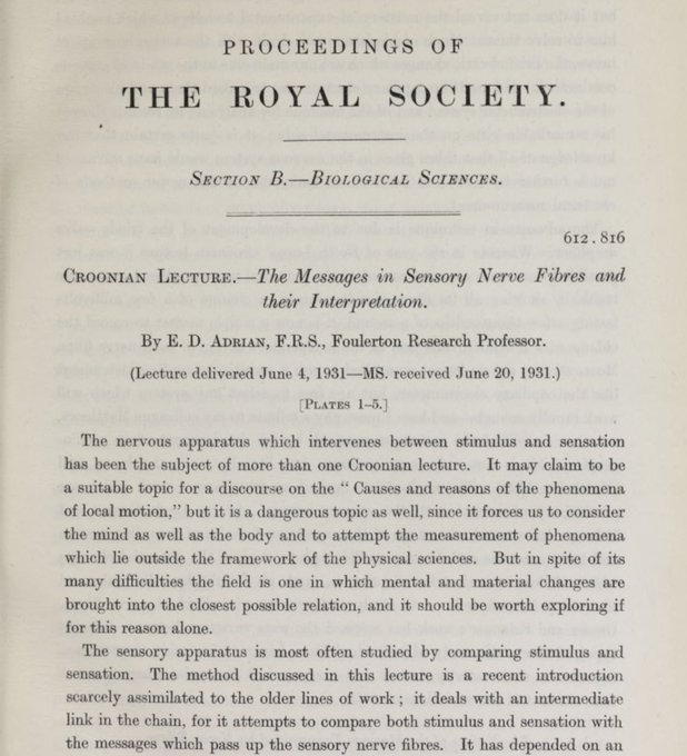
How are neurons activated by sensory stimuli? For the past century, the only method for addressing this question has been neurophysiology.
1
79
349
1
0
0
The above functional subsystems are speculative, but demonstrate the power of connectomics. We now have plausible guesses concerning the functions of all cell types intrinsic to the optic lobe!
1
0
0
Our guesses follow from Marsel Mesulam's maxim: “Nothing defines the function of a neuron better than its connections.”

1
0
0
To avoid overwhelming the viewer, all the wiring diagrams above show only the top input and output connections of each type, and types with relatively few neurons are also suppressed.
1
0
0
Here’s a single wiring diagram that includes all types intrinsic to the optic lobe, colored according to membership in the functional subsystems defined above.

1
0
2
Although the graph is highly sparsified for visualization (top input and output connections of each type only), there are prominent “hubs” (large symbols) like Tm1 and Mi1 that have many partners.
1
0
0
The new version of the paper has been upgraded from v630 to v783 of the reconstruction, increasing the number of proofread cells from 37k to 38.5k. Also 4700 photoreceptor cells have been added. Here's the v783 census.

1
0
1
We have clarified the hierarchy. Intrinsic neurons are divided into (a) columnar, (b) interneuron, (c) cross-neuropil tangential, and (c) cross-neuropil amacrine. Each class is divided into families, and each family into types.

1
0
1
We would like to congratulate our colleagues
@janeliaflyem
@MichaelBReiser
for releasing their optic lobe reconstructed from a male fly. We have been waiting to compare with our female optic lobe, and now we finally have the opportunity!

🧠Janelia scientists have reached another milestone in connectomics: A wiring diagram of the fruit fly visual system.
With 50K+ neurons, the optic lobe connectome provides the fullest picture yet of one of the most important parts of the nervous system.
0
74
278
1
5
21
@janeliaflyem
@MichaelBReiser
An obvious difference: the
@janeliaflyem
optic lobe has 900 medulla columns, while ours has 800. Could this be sexual dimorphism? Or is it just random variation between two individuals?
1
0
2