
SAR DFP Benign Biliary Pathology
@SARBiliaryDFP
Followers
364
Following
50
Media
48
Statuses
150
Official Account of the Society of Abdominal Radiology (SAR) Benign Biliary Pathology Disease Focused Panel
Joined March 2021
Stable fundal adenomyomatosis with endophytic component mimicking a polyp/mass
Biliary case from Dr. Karthik Sundaram and Dr. Matt Morgan at University of Pennsylvania, answer to come next week
0
0
1
Biliary case from Dr. Karthik Sundaram and Dr. Matt Morgan at University of Pennsylvania, answer to come next week
0
0
1
Next up, learn about benign biliary conditions with increased risk of malignant lesions @RachitaKhot @MayoRadiology
https://t.co/17wwPyOzkW
link.springer.com
Abdominal Radiology - Numerous conditions and pathologies affect the biliary system, many of which have underlying benign courses. However, these overall benign conditions can predispose the...
0
0
0
@MayoRadiology @ItaniMalak @UVARadiology @mghradchiefs @WyanneLaw @RachitaKhot @SocietyAbdRad @IURadiology @MDAndersonNews @PennRadiology @JenaDepetrisMD @WashUMedMIR Up next is a great review on acalculous cholecystitis! Check it out! @PennRadiology @RachitaKhot @MDAndersonNews
https://t.co/db7cRihH5R
link.springer.com
Abdominal Radiology - In this review, we highlight current understanding of the pathogenesis of acalculous cholecystitis, as well as its key imaging and clinical features. We also review what...
0
0
0
@MayoRadiology @ItaniMalak @UVARadiology @mghradchiefs @WyanneLaw @RachitaKhot @SocietyAbdRad @IURadiology Further your knowledge with this excellent article on Cystic lesions and their mimics involving intrahepatic bile ducts and the peribiliary space! @RachitaKhot @UVARadiology @MDAndersonNews @PennRadiology @JenaDepetrisMD @WashUMedMIR
https://t.co/vQVsNWIknB
link.springer.com
Abdominal Radiology - Biliary and peribiliary cystic lesions represent a diverse group of abnormalities, often discovered incidentally during imaging for unrelated conditions. These lesions,...
1
3
6
@MayoRadiology @ItaniMalak @UVARadiology @mghradchiefs @WyanneLaw @RachitaKhot @SocietyAbdRad @IURadiology Up next is an awesome review article on biliary complications of surgical procedures, covering bile leaks, strictures and more! https://t.co/RYv6GqvSNY
link.springer.com
Abdominal Radiology - Post-surgical biliary complications increase morbidity, mortality, and healthcare utilization. Early detection and management of biliary complications is thus of great...
1
3
8
@MayoRadiology @ItaniMalak @UVARadiology @mghradchiefs Next is an awesome review article on Imaging and management of complications post biliary-enteric anastomosis! @WyanneLaw @UVARadiology @RachitaKhot @SocietyAbdRad @IURadiology
https://t.co/mRNUnSqaHy
1
1
4
First up is a great review article titled: "Pancreatitis-related benign biliary strictures: a review of imaging findings and evolving endoscopic management" @MayoRadiology @ItaniMalak @UVARadiology @mghradchiefs
https://t.co/cc1NJNdqbM
1
0
2
Over the next few weeks, we'll be highlighting some great articles from our recently published collection of papers on "Complications and interventions in Benign Biliary Pathologies" @SocietyAbdRad
1
0
4
Challenge yourself with a case brought to you by Malak Itani, MD @ItaniMalak from @WashUMedMIR: 70M with RUQ pain x 1 week with nodular thickening of the GB wall, superimposed on changes of cholecystitis on initial CT, s/p cholecystostomy tube on MR.
3
3
7

Join us tomorrow for a high-impact, case-based webinar focused on the diagnostic and management challenges of benign biliary pathologies. Virtually tomorrow, June 10 from 4-5pm CT! Learn More + Register Now: https://t.co/XzoOevaHVe
#SAR
0
7
19
@RachitaKhot @matt_m0rgan @SocietyAbdRad @PennRadiology @UVARadiology @mghradchiefs @BWHRadEdu @KURadiology @UKy_RAD_RES @MayoRadiology @MayoMN_RadRes Featuring our great moderators and speakers! Registration: https://t.co/isd2yfBTk3
0
5
7
Join us next week for a great case based webinar- Mastering the Complexities of Benign Biliary Pathologies, on June 10th from 4-5pm EST! Moderated by our very own @RachitaKhot and @matt_m0rgan. Information and registration link below, You don't want to miss it! @SocietyAbdRad
1
3
7
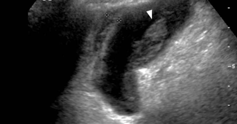
Axial and coronal contrast enhanced CT images demonstrating: Multiple discrete and confluent areas of cystic change within the gallbladder wall which is markedly thickened, continuous enhancement of the mucosa and serosa Answer: Xanthogranulomatous cholecystitis
0
1
4
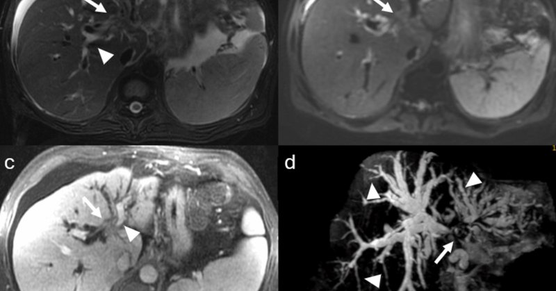
•Pre and post-contrast T1 fat saturation images: predominately T1 hypointense mass with areas of intrinsic enhancement. Answer: Intraductal papillary neoplasm of the bile duct (IPNB)
0
0
1
Axial diffusion weighted imaging: mass and filling defects demonstrate hyperintense signal on high B value diffusion imaging •MRCP and axial T2 fat saturation: diffuse intrahepatic biliary duct dilation; axial T2 fat saturation images confirm HASTE findings.
0
0
0
•Axial and coronal T2WI (HASTE sequences): An ~9cm hepatic mass with heterogenous, but predominately hyperintense signal, on T2WI. Associated intrahepatic biliary duct dilation with multiple curvilinear hypointense striations/filling defects.
0
0
0
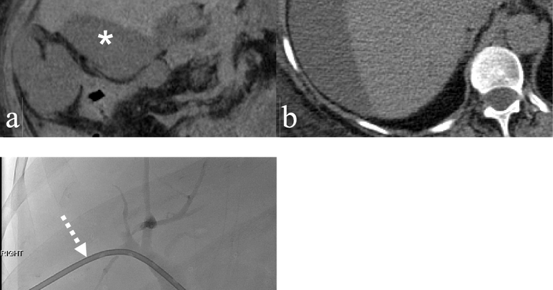
Challenge yourself with a biliary case: Adult woman with pancreatic adenocarcinoma undergoing restaging CT, reportedly asymptomatic
2
2
7