
MIPAR Image Analysis
@MIPAR_Software
Followers
174
Following
41
Media
196
Statuses
415
Revolutionary image analysis software, capable of identifying and measuring features from nearly any image one can capture.
Columbus, OH
Joined October 2015
MIPAR performs sub-pixel CD metrology using adaptive edge extraction to measure linewidth, pitch, and critical features in SEM images. Learn more at https://t.co/8LyQZ5AzPn
0
0
0
Within MIPAR you can construct a fully automated “recipe” mixing cleanup, FFT-normalization, convolution filters and ML-enabled segmentation — then export it via API into a Python pipeline. Learn more at https://t.co/ABGuSyjeF5
0
0
0
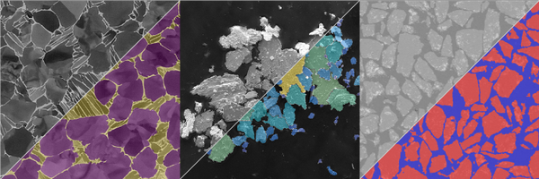
Automated boundary extraction quantifies grain size and morphology in multiphase alloys. Learn more at https://t.co/AnJfolzQQI
0
0
0
MIPAR automates radial unwrapping of Petri dish images for illumination-corrected colony segmentation and morphology-based enumeration. Learn more at https://t.co/SlrBbQessO
0
0
0
Automating microstructural workflows, MIPAR integrates deep learning segmentation with rule-based recipes for reproducible quantitative analysis. Learn more at https://t.co/Sfn41BfNBi
0
0
0
MIPAR quantifies pore size distribution by segmenting pores, extracting morphology metrics, and generating cumulative and differential distributions. Learn more at https://t.co/RrgS4jbnoO
0
0
0
MIPAR enables automated quantification of whole-slide pathology images, integrating deep learning segmentation with reproducible pipelines for histological feature extraction. Learn more at https://t.co/qVIXV76vxs
0
0
0
Quantify powder morphology with MIPAR: automated particle size and shape analysis yields reproducible metrics for distribution, aspect ratio, and sphericity. Learn more at https://t.co/nEdC9zGH3w
0
0
0
MIPAR automates phase segmentation in metallography, quantifying phase fractions and morphologies directly from micrographs. Learn more at https://t.co/CTljrM9YPu
0
0
0
MIPAR deep learning models segment contaminants across diverse substrates, enabling precise quantification of inclusion morphology and spatial distribution. Learn more at https://t.co/2ishlW4ox4
0
0
0
Automate segmentation of overlapping colonies in Petri dish images using thresholding, watershed, and circularity filters. Learn more at https://t.co/SlrBbQessO
0
0
0
Cell segmentation with MIPAR leverages local contrast and shape-based filtering to isolate overlapping, irregular, or faint cell boundaries in complex biological images. Learn more at https://t.co/NSJHZsv8dT
0
0
0
Quantify porosity and pore size distribution across scales using MIPAR's automated segmentation and measurement tools. Ideal for membrane, scaffold, and foam analysis. Learn more at https://t.co/duhwN2LmQT
0
0
0
AI-powered segmentation in MIPAR enables high-throughput phenotypic quantification of cell morphology across diverse imaging modalities. Learn more at https://t.co/nj04MYjUbr
0
0
0
Automated segmentation in MIPAR accelerates digital histology by quantifying structures in high-resolution slides with reproducible precision. Learn more at https://t.co/oQNyfsdnWc
0
0
0
MIPAR enables precise terrain classification and feature extraction from drone-captured orthomosaics using pixel-level segmentation and batch processing. Learn more at https://t.co/rboX2dWGQK
0
0
0
Die-to-die overlay error detection using MIPAR enables sub-micron misalignment quantification across wafers, supporting high-precision yield improvement. Learn more at https://t.co/xQawxfLsv4
0
0
0
MIPAR segments complex microstructures to quantify grain boundaries, inclusions, and phase areas in heterogeneous materials. Learn more at https://t.co/iMOpXhzs6E
0
0
0
MIPAR AI-aided thermal imaging segments rail defects, flags fires/obstacles from drone views, enhancing high-speed safety. Learn more at https://t.co/77OISYtA6y
0
0
0
MIPAR DL meets ASTM E112, auto-segments grains and outputs size distributions plus reports for metals and ceramics. Learn more at https://t.co/ML3kYvJwjH
1
0
1