
JournalofMicroscopy
@JofMicroscopy
Followers
4K
Following
731
Media
628
Statuses
3K
For scientists & technologists using any form of microscopy, the Journal publishes quality review articles and original research papers.
Oxford, UK
Joined September 2009
Don't forget - abstract submission and registration are *OPEN* for the 21st International Microscopy Congress #IMC21 🙌 🙂 🔬 (31 August - 4 September 2026) Be sure to take your place at the world's premier event for the global microscopy community! https://t.co/lFi1kOQWno
0
2
3
"Multiscale characterisation of cellulose nanofibril networks using three 3D imaging methods" by Nelly Vanessa Padilla Bello et al is available to view here:
onlinelibrary.wiley.com
Cellulose materials are suitable to replace plastic in food packaging. They are hydrophilic and may have poor barrier properties that affect the shelf life of the food due to the migration of...
0
0
0
🔬 In this new paper, the 3D microstructure of two different bilayer materials obtained with two Microfibrillated Cellulose (MFC) grades is investigated. For the first time, a full 3D representation of such material is presented.
1
0
0
The DAIM special issue can include invited manuscripts, original research articles, reviews, and methods & protocols. Deadline for submissions: 28 February 2026 More information can be found here:
rms.org.uk
0
0
0
The Guest Editors for the issue will be Rocco D'Antuono (The Francis Crick Institute - DAIM RMS, UK), Nicholas Condon (University of Queensland, Australia) and Beth Cimini (Broad Institute, US).
1
0
0
📣The Data Analysis in Imaging (DAIM) RMS science section and international collaborators have launched a special issue of the Journal of Microscopy, with the topic: “High-Performance Computing and Cloud-based solutions for image analysis in life and physical sciences”.
1
2
2
We are very pleased to announce that Professor Angus Kirkland, of @oxfordmaterials, @rosfrankinst & @diamondlightsource is to receive an RMS Honorary Fellowship - our highest accolade - for outstanding contributions to electron microscopy. Read more: https://t.co/Kj9XHZf4bv
0
2
4
This is well worth reading. An incredible amount of support for our community. Just incredible how much the @RoyalMicroSoc has delivered for us all in 2025, and this was before the vEuropean Flow meeting that attracted over 350 people from over 40 countries!

It's certainly been a busy year at the RMS - and you can read more about what we've been up to over the last 12 months in our special 'Annual Highlights' feature in the latest issue of infocus Magazine #RMSinfocus 🙌 Read more: https://t.co/5gejky6eoV
0
3
7
"Integrated approaches for multiscale mitochondrial structure and function analysis" by Adiba Patel, Prasanna Venkhatesh, Suraj Thapliyal, Margaret Mungai, Leo Jake Kazma, Muhammad Aftab, Antentor Hinton Jr, and Prasanna Katti is available to view here:

onlinelibrary.wiley.com
Mitochondria are double-membrane organelles whose architecture enables ATP (Adenosine Triphosphate) production, redox signalling, calcium homeostasis, and apoptosis. Visualisation of mitochondria...
0
0
0
They highlight the development of novel fluorescent probes, integrated correlative techniques, and computational analysis pipelines to expand the utility of mitochondrial imaging.
1
0
0
🔬 In this new review, the authors comprehensively examined the principles, capabilities, and limitations of diverse imaging modalities, with a focus on recent advances.
1
0
0
"A novel morphological descriptor for multiphase microstructure reconstruction" by DongDong Chen, JiaoFen Nan, XiaoRui Wang, XiangRu Chen and Bin Jiang is available to view here:

onlinelibrary.wiley.com
The microstructure distribution of multiphase porous media has important effects on its macroscopic properties. In this paper, we propose a framework for multiphase reconstruction based on entropy...
0
0
1
🔬 In this new paper, the authors propose a framework for multiphase reconstruction based on entropy statistical descriptor, which is used as morphological information to perform on two-dimensional (2D) and three-dimensional (3D) reconstruction.
1
2
2
📣There are more than 100 UK Institutions who are part of a deal between Wiley and Jisc which allows researchers free #OpenAccess publication in the Journal of Microscopy at no direct cost. Check your eligibility here > https://t.co/lQSsvLI8ta
@RoyalMicroSoc
0
1
4
"Comparison of different X-ray-based scanning electron microscopy methods to detect sub-nanometre ultra-thin InAs layers deposited on top of GaAs" by Thomas Walther, Stuart Creasey-Gray, Stephan Boehm, Heath Young and Yang Yang is free-to-view here:

onlinelibrary.wiley.com
Lay abstract: We compare three different methods of X-ray analysis in a scanning electron microscope (SEM): In energy-dispersive X-ray spectroscopy (EDX) the electron beam produces X-rays that are...
0
0
0
🔬 The authors of this new open access paper compare three different methods of X-ray analysis in a scanning electron microscope (SEM): energy-dispersive X-ray (EDX), wavelength-dispersive X-ray (WDX) and micro X-ray fluorescence (µXRF) spectroscopy.
1
0
0
📣The deadline for papers for the upcoming 'AI in Imaging' special issue has been extended to 31 December 2025. The special issue will be guest edited by Gianluca Tozzi, Peter Soar & Tuan Nguyen, all from the University of Greenwich. Read more here: https://t.co/OQx5Zjs2jz
0
1
2
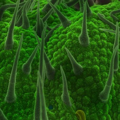
"Optical properties of cicada wings covered by graphene studied by nano-Raman spectroscopy" by Vitor A. F. Torres et al is open access and free-to-view here:

onlinelibrary.wiley.com
Some biological systems exhibit nanoscale constructions to produce optical effects. This study utilises Atomic Force Microscopy (AFM) and Tip-Enhanced Raman Spectroscopy (TERS) to study the complex...
0
1
1
The results demonstrated that light propagates preferentially between the nano-metric pillars, enabling chemical characterization of the biological surface and revealing both the graphene bands and the characteristic Raman peaks of the wing.
1
0
1
🔬By covering cicada wings with graphene, this study employed tip-enhanced Raman spectroscopy (TERS) combined with atomic force microscopy (AFM).
1
3
6