Gonzalo Serrano-Belmar.
@GSERRANOB_MSK
Followers
9K
Following
6K
Media
407
Statuses
1K
MSK radiologist at CLINICA ALEMANA SANTIAGO, Chile. Sharing knowledge in a didactic way. Amateur medical illustrator 🇨🇱+🇵🇹 #MSKRad
Santiago, Chile.
Joined March 2020
We frequently see on MRI inflammatory changes of the suprapatellar or prefemoral fat pads, and we call it impingement. But few times we see it directly as in these case. Impingement with painful snapping between both fat pads during knee flexion-extension. 💪 Dynamics . #mskrad
37
201
884
I am truly honored to be accepted as a new member of the International Skeletal Society. Joining this prestigious and inspiring community dedicated to musculoskeletal radiology is a great privilege. I would also like to extend my heartfelt gratitude to Dr. Cerezal and Dra.

7
0
43

Generate videos in just a few seconds. Try Grok Imagine, free for a limited time.
408
171
2K

Illustration/ultrasound anatomy. The flexor digitorum superficialis (FDS) and flexor digitorum profundus (FDP) tendons course within a common synovial sheath in the digits. At the level of the proximal phalanx, the FDS splits to allow the FDP to pass through and reach its
1
25
76
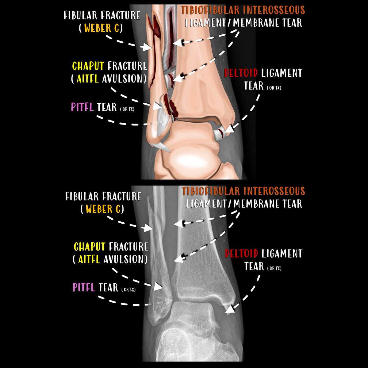
Mortise View – Stage 4 Pronation–External Rotation Injury. This mortise radiograph demonstrates a stage 4 pronation–external rotation (PER) ankle fracture-dislocation, following a predictable biomechanical sequence:.1.Medially, there is widening of the medial clear space,
0
4
21
An intrasubstantial tear of the Achilles tendon, especially in the context of tendinosis, represents a critical phase of fibrillar disorganization in an already degenerative tendon. On ultrasound, it presents as a poorly defined hypoechoic defect without obvious discontinuity or
1
20
122
Achilles tendon rupture most commonly occurs in the mid-substance region, approximately 2–6 cm proximal to the calcaneal insertion, where the tendon is relatively hypovascular and often shows underlying tendinosis. On ultrasound, a hypoechoic or anechoic gap may be seen
1
38
173
Proximal hamstring injuries. Ultrasound and MRI correlation demonstrates deep fiber avulsion at the proximal hamstring origin, with preservation of superficial components. The laminar hypoechoic cleft seen on ultrasound corresponds to a hyperintense interface on MRI,

2
22
109
Proximal hamstring injuries. It is common to observe a partial avulsion pattern where the deep tendon fibers detach from the ischial tuberosity while the superficial components, including extensions toward the sacrotuberous ligament, remain continuous. This anatomical
2
15
75
Multi-Imaging Correlation of Posterior Tibial Retinacular-Periosteal Malleolar Stripping. This case illustrates the value of multi-modality imaging—ultrasound, MRI, X-ray, and dynamic evaluation—in detecting posterior tibial tendon instability caused by retinacular-periosteal

3
17
78
Posterior tibial tendon instability due to retinacular injury. The flexor retinaculum is detached from its medial malleolar insertion, creating a retinaculo-periosteal pocket into which the posterior tibial tendon can dynamically dislocate. This instability may lead to chronic
5
27
130
Peroneal fracture, unstable fragment: Ultrasound versus MRI correlation.

0
12
68
Dynamic ultrasound revealed a non-union fracture of the distal fibular malleolus involving the insertion of the lateral collateral ligaments. Under stress, the fragment displaced, allowing intra-articular contrast to extravasate into a subcutaneous soft tissue pocket.
0
13
57
RT @DmcRadiologia: CIRME 2025 Santander, May 16–17. Multidisciplinary meeting on key topics in muscle injuries and upper extremity patholog….
0
19
0
RT @Rheumatology: Here is a variant to @GSERRANOB_MSK way to explain and show an in plane and out of plane injectio….
0
24
0

Las infiltraciones ecoguiadas pueden realizarse mediante dos técnicas principales: en plano (in-plane) y fuera de plano (out-of-plane). En la técnica en plano, la aguja avanza paralela al transductor, permitiendo visualizar todo su trayecto en tiempo real, lo que ofrece mayor
3
26
126