
Meghan Herbst
@EUSmkh
Followers
3K
Following
2K
Media
915
Statuses
2K
Ultrasound Enthusiast, Emergency Physician
Hartford, CT
Joined May 2013
Students: What organ is being viewed?.Residents: Which direction is the indicator pointing?.#raysofgray #POCUS #ultrasound #FOAMed #MedEd #emergencymedicine #medicalstudent #resident
1
0
4
RESOLUTION:.S: Phased array probe, screen indicator screen-left (non-cardiologist convention), with probe indicator erroneously directed to the patient’s left. R: McConnell’s sign, concerning for acute pulmonary embolism.
0
0
0
Paramedics can incorporate cardiac PoCUS after brief training among patients with chest pain in a moving ambulance! Kudos to Dr. Thorne et al on this project! .

0
1
1
Now available: a comprehensive resource for clinicians seeking to deepen their expertise in diagnostic and procedural ultrasound in acute care settings. #POCUS #EmergencyMedicine #UltrasoundEducation.
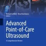
link.springer.com
0
0
1
Students: What probe is being used? Where is the indicator directed on the patient?.Residents: What is your interpretation?.#raysofgray #POCUS #ultrasound #FOAMed #MedEd #emergencymedicine #medicalstudent #resident
3
2
13
RESOLUTION.S: Convex probe over the LUQ of the abdomen. R: STOMACH. This pt had a gastric outlet obstruction from an incarcerated hiatal hernia.
0
1
0
Students: What probe is being used? Where do you think it is on the body?.Residents: What organ is being viewed? What is the pathology?.#raysofgray #POCUS #ultrasound #FOAMed #MedEd #emergencymedicine #medicalstudent #resident
1
1
3
RESOLUTION.S: Linear probe placed over the left lateral base of the neck, indicator directed anteriorly. R: A brachial plexus block somewhere between the interscalene and supraclavicular region of the neck is being performed to help with shoulder and arm pain.
0
3
3
Students: Where is the probe?.Residents: What procedure is being performed?.#raysofgray #POCUS #ultrasound #FOAMed #MedEd #emergencymedicine #medicalstudent #resident
1
1
5
RESOLUTION.S: A low-frequency convex probe is likely over the LLQ of the abdomen. R: Diverticulitis is the most likely diagnosis.
0
3
3
Students: What probe is being used, and where on the body is it most likely?.Residents: What diagnosis is consistent with this clip?.#raysofgray #POCUS #ultrasound #FOAMed #MedEd #emergencymedicine #medicalstudent #resident
1
2
3
RESOLUTION.S: Probe is over the abdomen.R: Small bowel obstruction: dilated (>2.5cm) small bowel (identified by plicae circulares) with alternating peristalsis. #raysofgray #POCUS #ultrasound #FOAMed #MedEd #emergencymedicine #medicalstudent #resident
0
2
2
Students: Where is the probe?.Residents: What diagnosis is most likely?.#raysofgray #POCUS #ultrasound #FOAMed #MedEd #emergencymedicine #medicalstudent #resident
2
5
7
RESOLUTION.S: Parasternal long-axis view with indicator screen-left convention (consistent convention for all applications).R: The ascending aorta is 6.5cm (normal <4 cm). In setting of chest pain, a type A dissection is the diagnosis of exclusion.
0
0
1
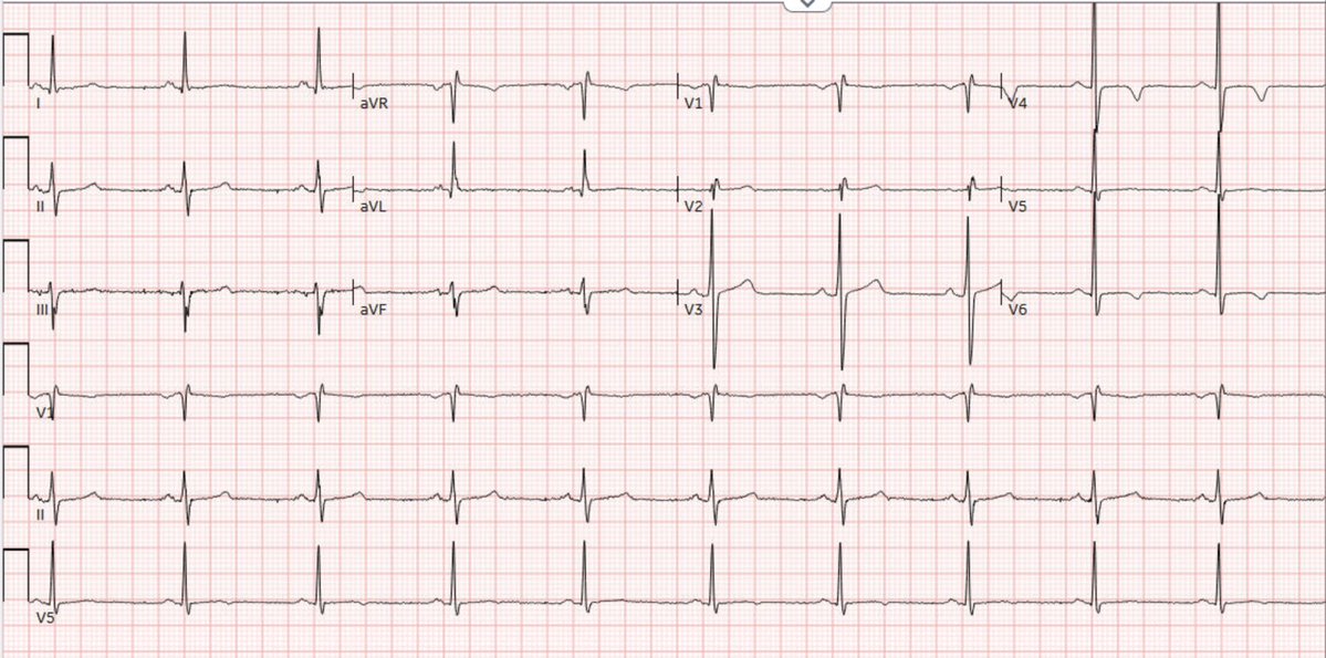
50-year-old male with chest pain radiating to jaw. No other symptoms. Students: What cardiac view is shown? .Residents: What diagnosis is most likely?.#raysofgray #POCUS #ultrasound #FOAMed #MedEd #emergencymedicine #medicalstudent #resident
2
0
7
RESOLUTION.S: Coronal plane, right pleural effusion .R: DDx—if bilateral: alveolar interstitial syndrome, fluid overload; if unilateral: pneumonia, pulmonary embolism, malignancy (hemothorax if trauma)
0
0
0
Students: What is the plane and finding shown? .Residents: What is your differential for this finding?.#raysofgray #POCUS #ultrasound #FOAMed #MedEd #emergencymedicine #medicalstudent #resident
1
1
2
RESOLUTION.S: Sagittal plane through the gallbladder (anterior to kidney, both long axis). Transverse would show both in short axis, & coronal could show one, but not both in the same plane. R: Common bile duct (CBD) dilation & biliary sludge.
0
1
1
Students: In what anatomical plane is this view obtained (transverse, sagittal, coronal)?.Residents: What pathology is shown?.#raysofgray #POCUS #ultrasound #FOAMed #MedEd #emergencymedicine #medicalstudent #resident
1
3
5