

CooperPathology
@CooperPathology
Followers
333
Following
22
Media
28
Statuses
53
Official Twitter account of the Cooper Pathology Residency Program at Cooper University Hospital @CooperHealthNJ
Camden, NJ
Joined January 2025
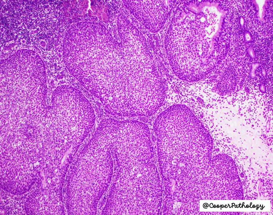
#pathology #pathtwitter Sinonasal Inverted Papilloma, a benign but locally aggressive tumor with endophytic growth. Presents as a nasal mass, often with obstruction or epistaxis. @navale_pooja @aileengraceA @ujoneja @324Amandeep @ruthbirbe @gord_zhu @dejan1295208

0
12
38
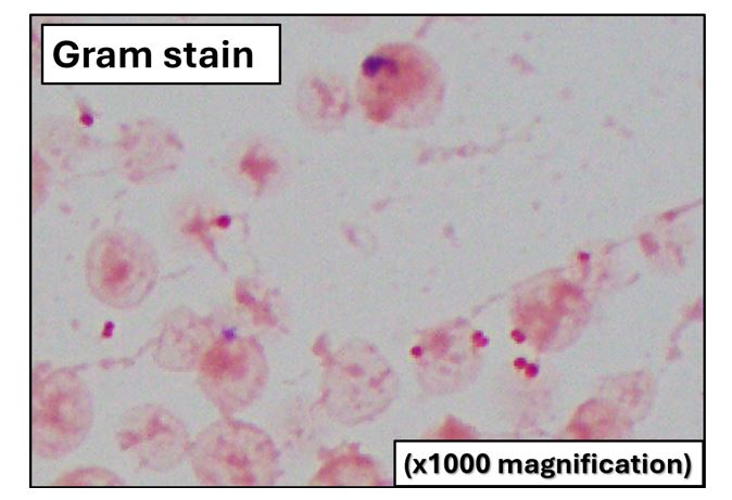
2/2 . Correct interpretation of the blood culture’s Gram stain?.A. Suboptimal Gram stain .B. Gram positive diplococci (indicative of Strep. pneumoniae).C. Gram negative diplococci (indicative of Neisseria spp.).D. Gram positive cocci (indicative of Staphylococcus/Streptococcus).
0
0
2
1/2 Micro alert! Case courtesy @dejan1295208.A young boy admitted to the PICU for acute onset fever, severe lower extremity myalgia, cool extremities, drop in BP, and change in mental status. Gram stain image of positive blood cultures shown below. Question on next page!
1
3
7
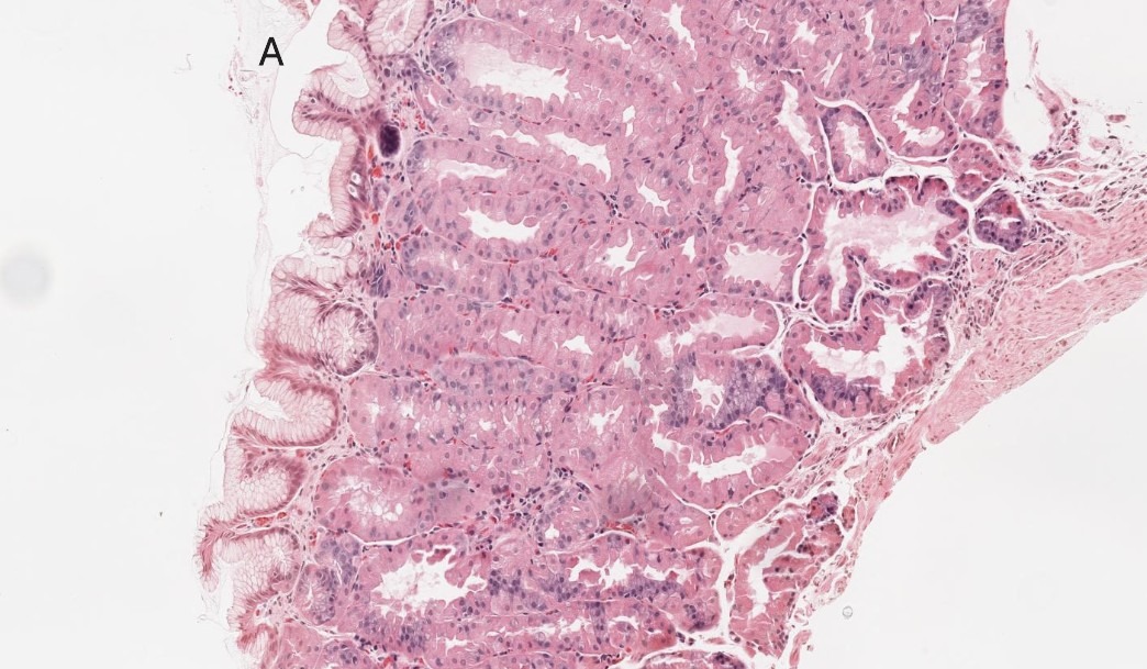
@AileenGraceA @navale_pooja @324Amandeep @Dejan1295208 @RuthBirbe @Gord_Zhu FGP: cystic dilated fundic glands .HP: elongated, hyperplastic foveolar epithelium, inflammation common, no dysplasia .FA: dysplastic foveolar cells, mimics HP but looks clonal, has nuclear atypia, neoplastic.PGA: small tubular glands, eosinophilic cytoplasm, looks clonal.
0
1
5
1
0
1
Stomach polyps #PathTwitter #pathresidents #pathology #stomach #polyps.A. Fundic gland polyp.B. Hyperplastic polyp.C. Foveolar adenoma.D. Pyloric gland adenoma.


2
11
54
1
13
34
Meet Dr. Upasana Joneja, MD @UJoneja -GI pathologist trained at Jefferson, Memorial Sloan Kettering, and Penn. She's passionate about advancing patient care, educating trainees, improving field efficiency, and shaping the future of the field. #FacultySpotlight #Pathtwitter

0
3
10
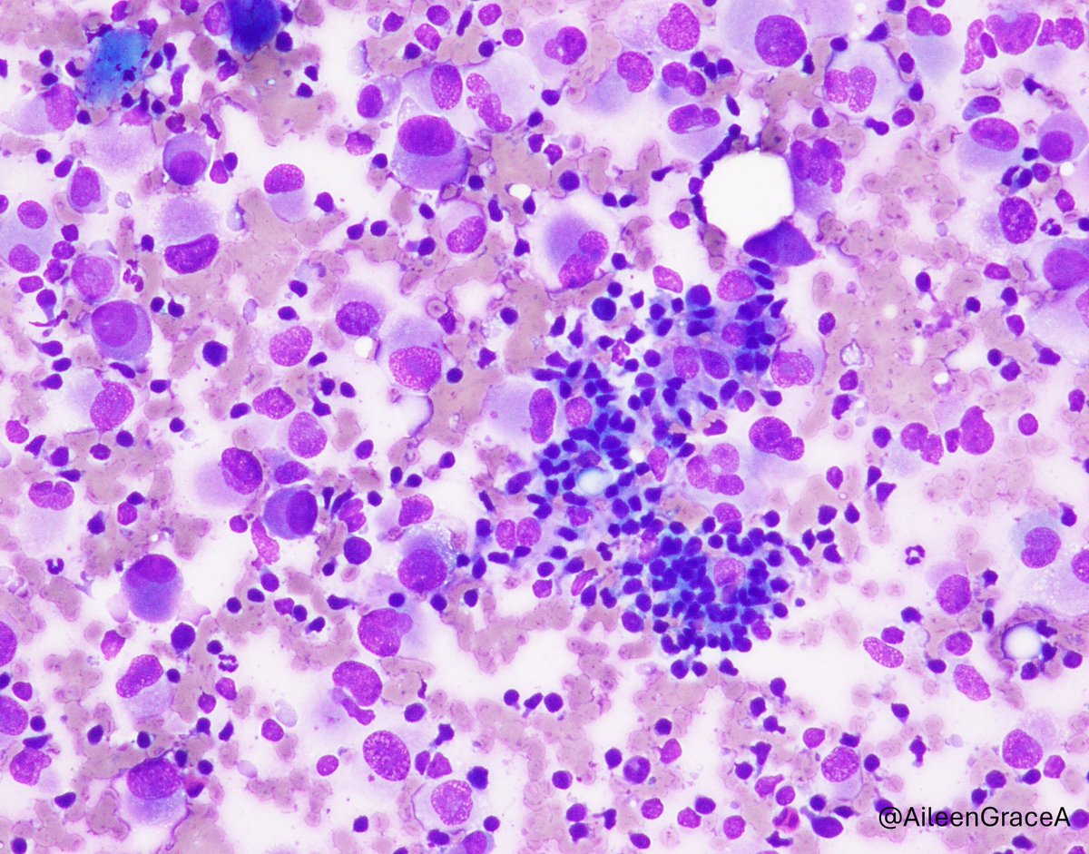
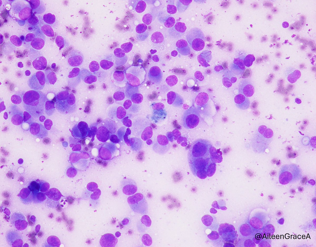
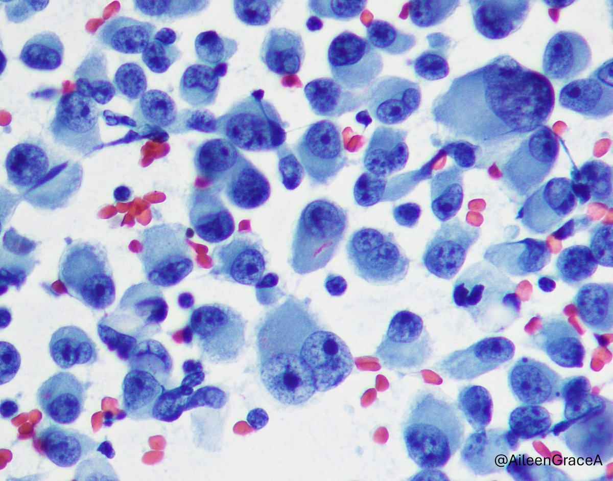
Kudos to @md_terzioglu and @Venkateshgilly2 for nailing this #cytopath case. This is indeed metastatic melanoma to a lymph node. There are large plasmacytoid cells with prominent nucleoli & occasional binucleation. Smaller cells are lymphocytes, lymphoglandular bodies in DQ.
Calling #cytopath enthusiasts. Can you name the diagnosis and site sampled based on these images?. #FNAFriday #PathTwitter #PathX

0
2
4
Calling #cytopath enthusiasts. Can you name the diagnosis and site sampled based on these images?. #FNAFriday #PathTwitter #PathX
4
8
17
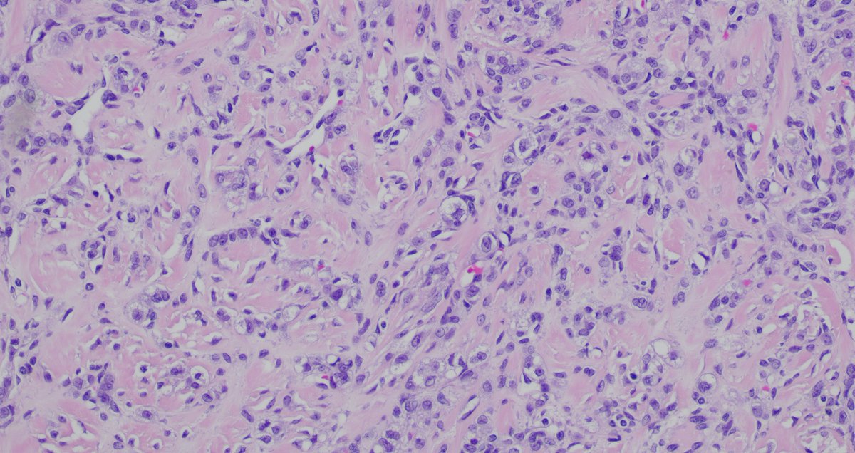
Final diagnosis is extra-axial chordoma. Here is the immunostain for Brachyury. @navale-pooja.@aileengraceA.@ujoneja.@324Amandeep.@ruthbirbe.@dejan1295208.@gord_zhu

1
2
8
0
0
1
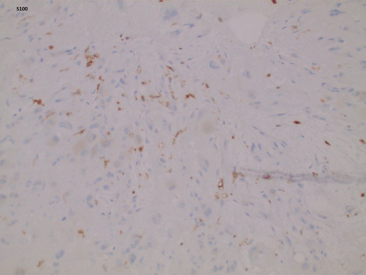
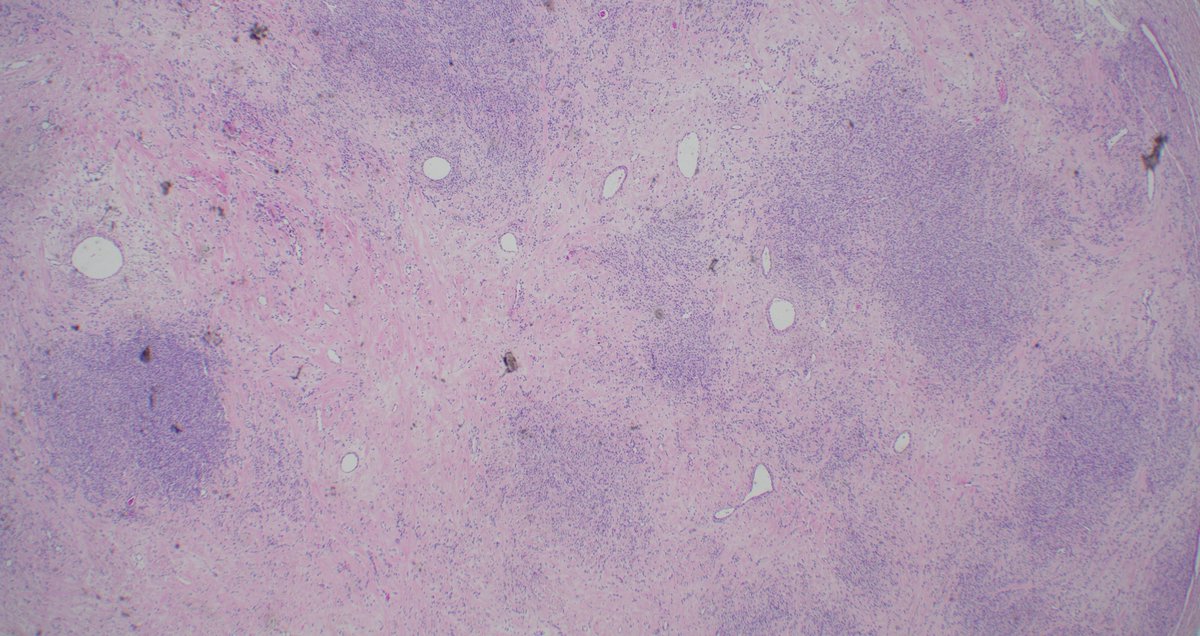
IHC profile: Strongly positive: EMA, CK19; focally positive for AE1/AE3, Cam5.2 and S100; Negative for CD31, CD34, SOX10, desmin and H3K36M. What additional stain do you want?


3
1
2


32 yo female presented with a knee mass. What are your ddx and workups?


4
6
14
Lactating adenoma of the Breast: benign, well-circumscribed mass typically seen in pregnancy/lactation. Histology: lobular expansion, secretory change, minimal atypia. Remember: can mimic carcinoma clinically/radiologically!.#PathTwitter #Breastpath


0
18
49
@navale_pooja @RuthBirbe @UJoneja @AileenGraceA @324Amandeep Great answers! Pseudolobulation, prominent ectatic vessels, and admixed spindled and luteinised cells give away the diagnosis - Sclerosing Stromal Tumor. Devins KM, Young RH, Watkins JC. Sclerosing stromal tumour: a clinicopathological study of 100 cases. PMID: 34467561.
0
1
5
1
0
0
A 32-year-old female with a 4 cm left ovarian mass. What is your diagnosis? #PathTwitter #pathology


9
19
60
Honored to be led by Dr. Roland Schwarting, Chair of Pathology at Cooper & Professor at CMSRU. A hematopathology expert trained at Kiel & Sloan Kettering, his vision, mentorship & patented tumor marker innovations(CD22, CD30, CD11c) continue to shape our department #PathTwitter

2
5
11