
Microscopy Chicago
@ChicagoMicro
Followers
181
Following
52
Media
33
Statuses
2K
Microscopy in Chicago
Chicago, Illinois
Joined June 2020

The Nikon AX / AX R #Confocal #Microscopy system and silicone immersion objective lenses – a decisive combination for imaging thick samples such as tissues and #Organoids with minimal spherical aberration. Click to learn more about objectives for the AX: https://t.co/necRtdsE6Z
0
4
15
#ImageScanningMicroscopy #ISM has arrived at Nikon! Introducing NSPARC, a SPAD array-based detector for the Nikon AX / AX R #Confocal #Microscopy systems. NSPARC provides #SuperResolution and with superior sensitivity compared to conventional GaAsP PMTs: https://t.co/JXGQ1oB8Ah
0
16
48
Did you know that you can try our Denoise_ai #DeepLearning-based denoising software for free on your #Confocal #Microscopy images? Click to check it out: https://t.co/82L09HEsJj
0
2
8
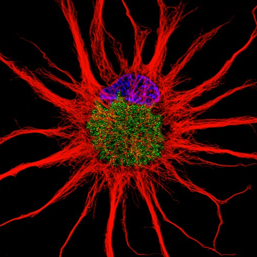
New #NikonSmallWorld Masters of Microscopy interview featuring Dr. Eduardo E. Zattara – winner of this past year’s @NikonSmallWorld video #Microscopy contest for his video of sensory organ progenitors (green) migrating along the lateral line in #Zebrafish: https://t.co/I09GxZNcoM
0
4
14

超解像共焦点レーザー顕微鏡システム 「AX/AX R with NSPARC」なら、超解像によるサンプルの詳細な微細構造の取得・測定・解析までが可能になります。 ▼製品の詳細はこちら https://t.co/SVpsZd2Don
#Nikon #顕微鏡 #新製品
0
3
15
This week's #AXReview comes from Dr. Paolo Bianchini @2Pbianchini, the Facility Manager of the Nikon Imaging Center at @IITalk. Click for more info about the AX #Confocal #Microscopy system: https://t.co/jFE8vqfKmY
0
4
15

And now, the stereo microscope SMZ18 @NikonInst has a new sCMOS ORCA Camera for High Speed Large FOV imaging...stay tuned to see what we do with it 😉😎
0
3
36

And finally, the winner of the Images of Research competition sponsored by @NikonInst was Karla Neves with Blood Vessel Comet! Congratulations @kbneves!
0
2
21
Thank you again to everyone who entered the competition, you can find more about upcoming competitions and the society at: https://t.co/pQqp61W2ZO Thank you to all our sponsers @VisiTech_UK, @focalplane_jcs, @NikonInst and @zeiss_micro!
0
1
6
Ammonites, which evolved about 416 million years ago, were once the most abundant animals of the ancient seas. https://t.co/grvGrVjBVX Photo Cred: Dr. Balint Markus
0
3
12
Cell division is how animals grow and reproduce. Each new cell has the same genetic material (DNA) as the cell that produced it. https://t.co/I0smKK67ud Photo Cred: Dr. Andrew Moore & Dr. Erika Holzbaur
0
5
30
You have until 5pm today to enter the @scotmicro Christmas Image Competition, < 6hrs to go! Enter here: https://t.co/5IpMfxjUfX… Thanks to our sponsors @NikonInst @focalplane_jcs @zeiss_micro @VisiTech_UK £50 prize up for grabs in each category! 🥳 #microscopy #imaging
1
8
10
The @scotmicro Christmas Image Competition closes this Friday! Enter here: https://t.co/5IpMfx1L1P… Thanks to our sponsors @NikonInst @focalplane_jcs @zeiss_micro @VisiTech_UK £50 prize up for grabs in each category! 🥳 Closes 5pm, 9th Dec Please RT 🙂 #microscopy #imaging
0
12
14

How cool is that? Our yeast mitochondria on the ceiling of The National Museum of Natural History by @NikonInst Amazing @felix_thoma !!

0
5
39

We are ready! Booth 92O #Cellbio2022 displays the X-Light V3 #spinningdisk / #DeepSIM Super Resolution System on a Ti2E inverted platform in collaboration with @NikonInst @Photometrics @HamamatsuPhoton Meeting you all is going to be incredibly fun! @ASCBiology
0
2
13
Thank you @NikonInst for launching CrestOptics #DeepSIM Super-Resolution system integrated with X-Light V3 #spinningdisk on a Nikon Ti2E Inverted Platform and NIS Elements. #Cellbio2022
@ASCBiology
0
3
10

Let's walk through a bioprinted channel of our vascularized #HUMIMIC Chip imaged via confocal microscopy by @NikonInst BioImaging Lab, Japan Green: F-Aktin, Endothelial cells Blue: DAPI, cell nuclei Red: Celltracker CMPTX, white blood cells (fixed) #MPS #organonachip #microscopy
0
4
13
That night I watched EB1 comets & cells divide in zebrafish until ☀️rise @MBLScience #embryo2017 #ThrowbackThursday #TBT #NikonInst #NikonA1
0
6
34