
Carol N. Rizkalla, MD
@Carol2Path
Followers
2K
Following
7K
Media
248
Statuses
1K
PGY4 AP/CP @UWlabmedpath 🇺🇸 26-27 @StanfordPath #GYNpath fellow 😍 @RCSI_irl 22 alum 🇮🇪 Creator of #PathWebinarPearls 🧵I Trainee #PathTweetAward 23&24
Seattle, WA
Joined September 2020
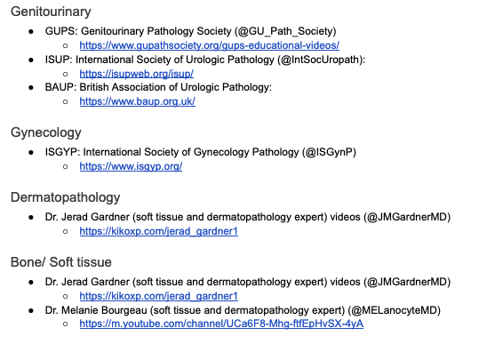
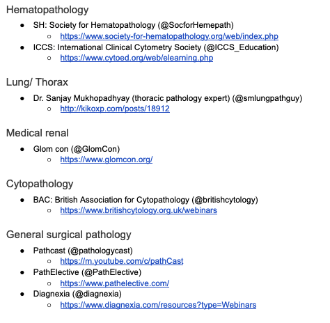
I am a huge proponent of educational webinars for learning 🧠 #PathWebinarPearls. 👇👇👇is a compiled list of societies that regularly host educational webinars- many of which I have listened to myself! Most of these are FREE for residents, you may need to register as a member.
8
94
211
#USCAP2025 w/ Dr Matias-Giu. In endometrial carcinoma, cells at the invasive front undergo EMT transiently and revert to an epithelial phenotype after invasion. In contrast, in carcinosarcoma, EMT is thought to be permanent rather than transient phenomenon . #PathWebinarPearls


3
11
45

Generate videos in just a few seconds. Try Grok Imagine, free for a limited time.
495
859
4K
RT @Janiranavarro: Morphologic correlation with molecular findings.66 yo male with a groin mass.This lesion stains with STAT6 (staining in….
0
26
0
#USCAP2025 w/ Dr Mete. Differentiated neuroendocrine neoplasms have lineage specificity- they replicate some of the developmental biomarkers based on their anatomic region or location. Contrast to NECs which ❌ respect lineage specificity (aberrant expression). #PathWebinarPearls

0
15
48


0
17
0
RT @Janiranavarro: “Everyday” Cutaneous Soft Tissue Tumors.How much keratin immunoreactivity do we need to make the diagnosis of sarcomatoi….
0
7
0

A select panel of histiocytic/osteoblastic/osteoclastic markers may be helpful in deciphering plasmacytoid UC from UCOGC. PMID: 40512053. NGS may also be helpful as plasmacytoid UC classically harbors 🧬CDH1 mutations🧬.

1
0
1
All 14 cases in our series had conventional UCOGC areas + a mononuclear component resembling plasmacytoid UC. Interestingly, 30% of the tumors also showed true plasmacytoid UC morphology as a distinct subtype

1
0
0
🚨 Morphologic and immunophenotypic overlap🚨. Urothelial carcinomas w/ osteoclast-like giant cells (UCOGC) can have mononuclear cells w/ eccentric nuclei resembling plasmacytoid UC. These cells also show loss of E-cad & cytoplasmic p120 staining.

1
11
28
#USCAP2025 w/ Dr Voltaggio. Identifying 4 lines helps avoid missed dysplasia in upper GI & reduces equivocal cases → fewer interventions. Loss of lines = dysplasia. 4 Lines:.1.mucin cap.2.bottom of mucin cap.3.cytoplasm.4.nuclei. #PathWebinarPearls


1
47
143

#USCAP2025 w/ @JLHornick . Undifferentiated and dedifferentiated melanomas are tough 😨, they have lost staining for conventional melanoma markers. Using mutation-specific IHC BRAF V600E and considering clinical history (ie. axilla) are key for diagnosis. #PathWebinarPearls


0
31
61
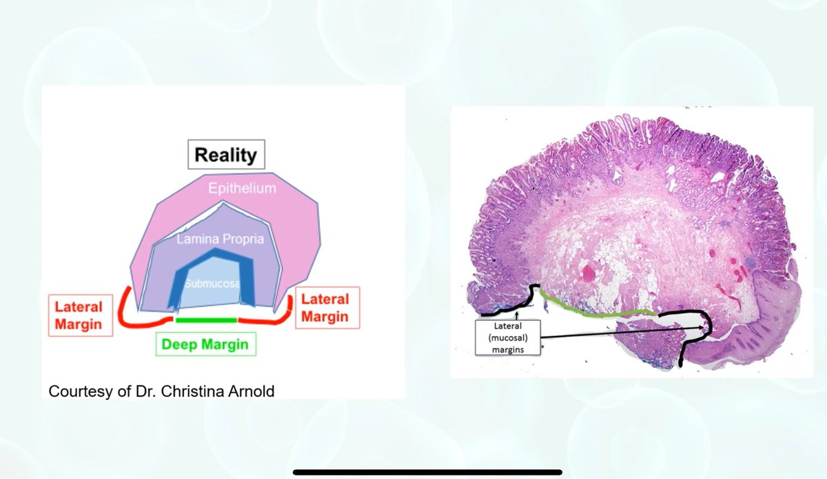
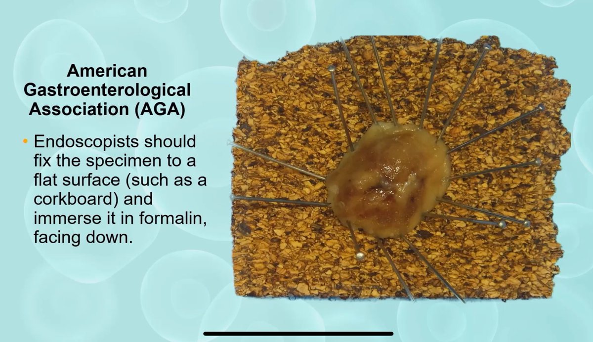
#USCAP2025 w/ Dr Voltaggio. Esophageal ⬆️ grade dysplasia/intramucosal adeno → treated with EMR. Presence of muscularis mucosa = lateral margin; large vessels + loose CT = deep margin. Recommendation to pin specimen before fixing for easier margin assessment. #PathWebinarPearls

2
39
93
RT @RazaHoda: No doubt—you all crushed it! 💯. The answer: B-cell lymphoma. This one’s a marginal zone lymphoma of the breast—rare, and a g….
0
6
0
RT @TingZhaoPathdoc: 30s male with renal mass. Outside biopsy dx: “RCC, favor chromophobe” (photos below). Repeat biopsy + nephrectomy phot….
0
23
0
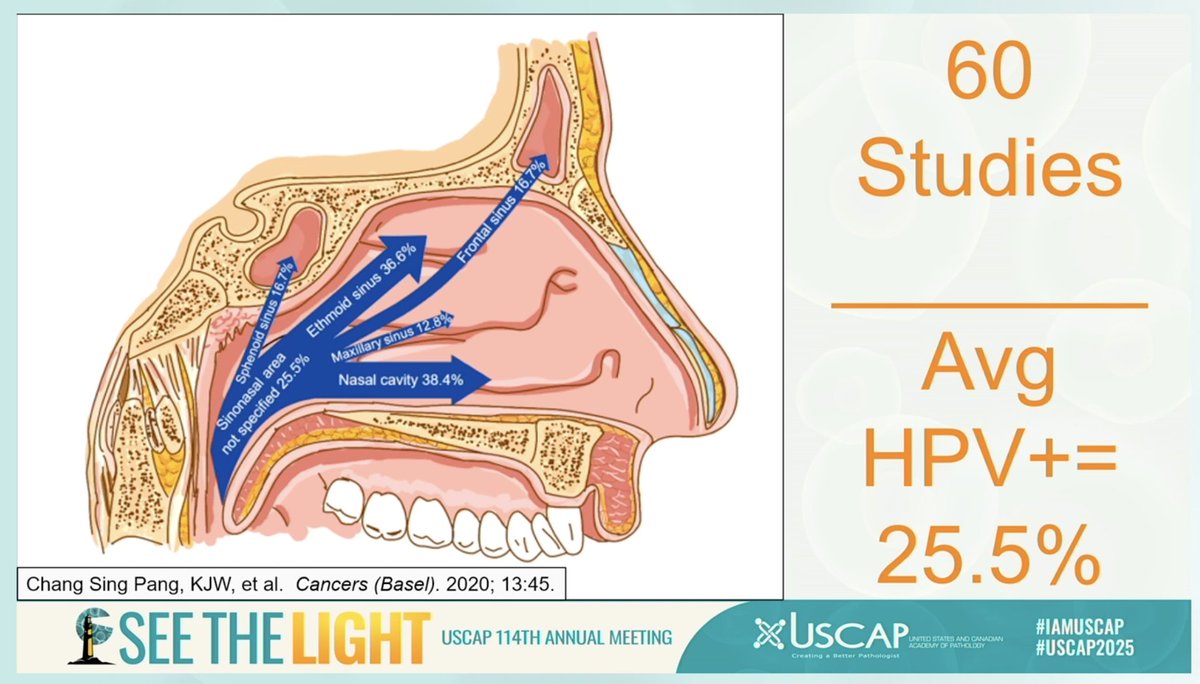
#USCAP with Dr James Lewis . We know to test all oropharyngeal cancers for HPV but what about ✨sinonasal✨? --> recommendation to also test these for HPV (25% associated w/HPV). Mechanism? pharyngeal reflux, so still thought to be sexually transmitted. #PathWebinarPearls


0
7
27
RT @_TaraKrishnan_: Recently I gave a talk at our @UWLMPathRes Research Rounds about improving surgical specimen workflows via a combined s….
0
1
0
RT @_TaraKrishnan_: Thank you for an amazing lecture and lunch session @KMirza ! Dr. Mirza spoke about personalized education📝 in the web-b….
0
2
0
RT @KMirza: A spectacular day in Seattle visiting the @UWLMPathRes! Thank you for your incredible hospitality and the inspiring conversatio….
0
4
0
RT @Janiranavarro: Leiomyosarcomas in soft tissue-core biopsies. -Any mitotic activity, atypia or necrosis is enough to call it leiomyosarc….
0
23
0