
BIGR
@BIGR_ErasmusMC
Followers
206
Following
25
Media
10
Statuses
201
Biomedical Imaging Group Rotterdam | The Netherlands | Erasmus MC @[email protected]
Rotterdam
Joined October 2020
RT @EuroBioImaging: At the #EuroBIoImaging User Forum, Jukka Hirvasniemi, @BIGR_ErasmusMC, presents the KNee OsteoArthritis Prediction (KNO….
0
1
0
RT @EuroBioImaging: Our Population Imaging Node @BIGR_ErasmusMC contributed to the recent #EUCAIM platform release - an important step tow….
0
4
0
Recently the new ICAI Stroke lab has started at @ErasmusMC and @erasmusuni. Five PhD students, including one in our group, will work on improving outcome for stroke patients using AI techniques, in collaboration with @ic4ai and @PhilipsHealth
amazingerasmusmc.nl
In het pas opgerichte ICAI Stroke Lab gaan vijf promovendi zich onder begeleiding van een team van experts van het Erasmus MC en de Erasmus Universiteit in samenwerking met Philips richten op het...
0
1
7
The title is "Automated Deep Learning–Based Segmentation of Abdominal Adipose Tissue on Dixon MRI in Adolescents: A Prospective Population-Based Study" and the paper can be found here:

ajronline.org
BACKGROUND. The prevalence of childhood obesity has increased significantly worldwide, highlighting a need for accurate noninvasive quantification of body fat distribution in children. OBJECTIVE. The...
1
1
2
A new paper by Tong Wu and colleagues was just published! Here, Tong presents the work on Automated Deep Learning–Based Segmentation of Abdominal Adipose Tissue: #imageanalysis #abdomen #medicalimaging #deeplearning @ARRS_Radiology.
1
1
9
RT @oliviercolliot: "Machine Learning for Brain Disorders" is now published! 1058 pages fully open access

link.springer.com
0
46
0
RT @esther_bron: Book out now on Machine Learning for Brain Disorders. Great effort led by @oliviercolliot to which we contributed a chapte….
0
2
0
Open position for a PhD student on muskuloskeletal image analysis: We are looking for a PhD student to develop and apply #deeplearning-based #imageanalysis techniques for assessment of hip #MRI scans of young adults in a large GenerationR study.
0
1
1
New position open for the Erasmus MC Imaging Office. We are looking for a processcoordinator/developer that willl coordinate and develop services related to medical imaging
0
1
8
RT @midl_conference: Here’s a glimpse of the first @ViseVanderbilt tour of the Conference! Be sure to follow VISE on social media to stay i….
0
2
0
Congratulations to Stefan Klein and @MartijnStarmans for their grant on the iver #ArtificialIntelligence (LAI) Consortium!.

👏Congratulations to dr. Stefan Klein with his @NWONieuws #OpenTechnology #grant for his #project "The Liver #ArtificialIntelligence (LAI) Consortium: A Benchmark Dataset and Optimized #MachineLearning Methods for #MRI based Diagnosis of Solid Appearing #Liver Lesions". [1/2].
0
1
8
Now funded & to start soon: the Liver Artificial Intelligence (LAI) consortium! @MartijnStarmans and colleagues aim to build the biggest publicly available (imaging) benchmark for liver AI and a clinical validation AI solution to aid in liver tumor phenotyping.
Very happy to announce that Stefan Klein, Maarten Thomeer, and myself got a @NWONieuws Open Technology grant on our Liver Artificial Intelligence (LAI) consortium:
0
1
7
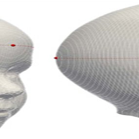
New article by Tareq Abdel Alim and colleagues: Reliability and Agreement of Automated Head Measurements From 3-Dimensional Photogrammetry in Young Children @JCSOnline1
journals.lww.com
3D images of 188 patients diagnosed with sagittal synostosis using a novel automated method proposed in this study. In addition, the study aimed to determine the interrater and intrarater reliability...
0
1
3
High quality Research Software & Research Software Engineers are very crucial to our research! #researchsofware.

📢 The @esciencecenter released its RSE job profile & role description to bring recognition to #researchsoftware & RSEs! They can serve as resources for those looking to define & appropriately position RSEs w/in their org. #RecognitionRewards. 👉 Please RT.
1
2
3
RT @EuroBioImaging: Great webinar on Data Management of Preclinical Image Datasets organised by @unito @CNRsocial_ in collaboration with th….
0
3
0
New paper by @daniloajesus and colleagues on retinal OCT speckle as biomarker for glaucoma diagnosis and staging
0
0
5

How quantitative is perfusion DSA? In this new paper @RuishengSu and colleagues investigated the quantitative properties of deconvolution-based #perfusion #DSA using animal imaging data. Now online in Medical Physics #MedPhys:.

aapm.onlinelibrary.wiley.com
Background X-ray digital subtraction angiography (DSA) is the imaging modality for peri-procedural guidance and treatment evaluation in (neuro-) vascular interventions. Perfusion image construction...
1
3
4