Albany Med Radiology Residency
@AlbanyMedRadRes
Followers
1K
Following
399
Media
43
Statuses
175
#RadRes of Albany Medical Center https://t.co/x3tTzDEtHG
Albany, NY
Joined March 2019
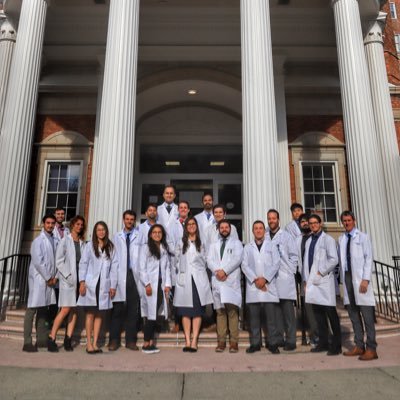
Warmest of welcomes and congratulations to our newly matched Albany Med DR and IR residents 👏🏻 👏🏻 @AlbanyIR #Match2024 #FutureRadRes
0
5
29
@AlbanyMedRadRes came out strong 💪 this morning at Saratoga State Park for the annual #SarcomaStrong 5k. VIR resident Jonathan Long raised $1260 for #prayersforbehrs 🤩. Well done!! 👏🏻 👏🏻 @AlbanyMed @AlbanyIR
0
3
14
Joining the Twitter party to congratulate our current DR and IR residents who all successfully passed the CORE 💪. Well done Ted, John, Mai, Kris and Pagah! 👏🏻 🎈 🎉
0
1
20
Cannot thank @AlbanyMedRadRes @avnarayan @JosephGiampaDO and many more not on Twitter enough for the incredible training, mentorship, and friendship I have received throughout my residency! I will always keep in touch! Next stop, neuroradiology fellowship @PennRadiology 🧠
7
3
58

Congrats on residency graduation! @daniel_gewolb the neuro section @AlbanyMedRadRes will miss you! Looking forward to following more of your awesome cases at @PennRadiology
0
3
19
Belated thank you to Albany Medical College alum @nkagetsu for joining us last week at @AlbanyMed to discuss Unconscious Bias and Microaggression. Wonderful talk and wonderful discussion. @AlbanyIR @AlbanyMedRadRes
0
1
10
Thrilled for 3 of our trainees to be recognized for @AlbanyMed Resident/Fellow Recognition Awards. Congrats Ted Nicolosi (DR), RK Patel @ramswishna (Combined IR) and Anthony Sayegh (Independent IR). Well deserved👏🏻 👏🏻 👏🏻 @AlbanyIR @allen_herr @gsiskin #radres
0
1
12
Welcome to the team! Congratulations 👏🏻 👏🏻 👏🏻
2
7
40

This future #neurorad deserves more followers -- check out these high yield and practical teaching posts #radres 👇

CTA tip: Use the post con source images to evaluate the brain parenchyma. Here is an example of a metastatic lesion that was invisible on the unenhanced CT but incidentally found on the post con images (yellow arrow) #radtwitter @TheASNR #radiology
1
3
22
73 y/o M presents with palatal myoclonus. MR shows hyperintense and enlarged inferior olivary nuclei. #MedTwitter #radtwitter #radres #Neurology #Medstudent @ASNRographics
5
21
88
Child presents with weakness. MR shows enhancement of the pial surface of the conus and ventral cauda equina nerve roots. #radtwitter #MedTwitter #radres #futureradres #Pediatrics #Neurology @TheASNR @The_ASPNR @AlbanyMedRadRes
1
3
10
68 y/o asymptomatic patient presents with bilateral ill defined soft tissue masses with trace interspersed fat located in the infrascapular regions (yellow arrows) #radtwitter #radres #futreradres #medstudent
2
2
15
Diagnosis: Optic nerve sheath meningioma Remember the optic nerve is an extension of the CNS and therefore, is surrounded by meninges and arachnoid cap cells from which meningiomas arise. Look for the “tram-track” sign of enhancement surrounding the optic nerve #Ophthalmology
1
2
12
45F presents with painless progressive left eye vision loss. MR shows homogenous enhancement encasing the left optic nerve with an associated lesion at the entrance of the optic canal (yellow arrow) #radres #futureradres #NeuroRad #MedTwitter @AlbanyMedRadRes
5
11
45
62 y/o M presents with signs of raised intracranial pressure. CT shows a hyper dense mass crossing the corpus callosum. On MR, the mass is hypointense on T2WI, restricting diffusion, and homogenously enhancing along the periventricular surface. #radtwitter #radres #neurotwitter
3
15
47
Patient with leukemia presents with fever and nasal congestion. MR shows mucosal thickening with areas of absent mucosal enhancement (yellow arrows). DWI and post con of the brain show extra-axial restricted diffusion and enhancement. #radres #MRI #radtwitter @ASHNRSociety
3
25
61
The germinal matrix is a fragile capillary network that’s sits between the caudate nucleus and ependymal lining of the frontal horn of the lateral ventricles. The matrix resolves by 35-36 weeks gestation but is a common location of hemorrhage in premature infants. @The_ASPNR
1
5
12
78 yr old with encephalopathy and seizure. MRI shows vasogenic edema within the left temporal and occipital white matter with subcortical extension. SWI shows characteristic lobar micro-hemorrhages predominantly in the effected regions. #Neurology #neuroradiology #radres
1
2
6