
linda johnston
@oreoimc
Followers
2K
Following
13K
Media
2K
Statuses
5K
Cytotech -pathology /science nerd with a healthy spattering of sarcasm ( oh, and a proud Canadian🇨🇦)
Moncton, New Brunswick
Joined August 2014
A vulture ,a lizard,an elephant and a seahorse. A busy day in the Cytology Zoo
11
71
244

A cell block preparation from a lung nodule FNA in an older man with history of multiple myeloma. Notice the variably sized structures in the H&E stain, which are highlighted by GMS stain. Mucicarmine stain demonstrates the mucin capsule. Diagnosis? #CytoPath
7
19
52


22
554
3K

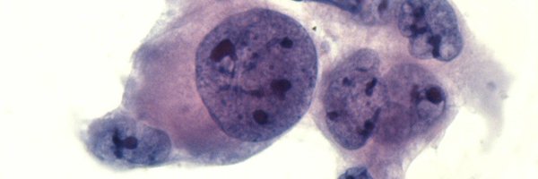
FNA of a cervical lymph node in a woman with a history of breast carcinoma of apocrine type.
3
17
48
@SamKhader @MyCytopathology @UPMCPath @UPMCPathology @PathPro @EHJPathDiva @DrEstherAdler @SaeedAsiry @Quiroga_GM @mreyesm What perfect descriptions! Great images as well 👏👏
0
1
2

Dr. Zubair Baloch (@aakasharmand) explaining a helpful cytologic clue to reactive pneumocytes in acute/subacute lung injuries. Their resemblance to Napoleon Bonaparte's iconic hat. Initially described by @natasharekhtman 🔗 to the article👇: https://t.co/XiuvQ8GPyI
#IACTutorial
2
16
56

2
27
82

✨COLLAGEN BALLS ✨ it is not uncommon to see them in #cytology samples from peritoneal washings, together with large sheets of benign mesothelial cells, perhaps a little angry..
6
34
74
causes of dilated cardiomyopathy find remaining here 👉👉 https://t.co/Jl40Emyp2X ... .... #MedEd #FOAMed #HeartDisease
@oreoimc @drmaypole @VijaySelvarajMD @Wilkinsonjonny @renalpages @KoraAbhi @padmapathology1 @physicianswkly
0
7
20

Thoracic masses. Can you guess how many immunostains were done and why? #pathology #pulmpath
18
47
174

What fungal infection is seen in this biopsy of the small intestine of an immunocompromised patient? Answer & additional stain: https://t.co/Aw4mx0VVcP
#PathArt #PathTwitter #IDTwitter #MedTwitter
12
71
263
0
25
63
Pleomorphic carcinoma, lung: Note: this is the thoracic WHO term for sarcomatoid carcinoma. As usual, they threw out a good term that everyone understands for awful, inane terminology.This case also... https://t.co/7admmThTsJ
6
45
199
Interesting morphology of a squamous cell carcinoma metastatic to the pleural cavity (not common to see SqCC in an effusion #CytoPath specimen). Some of the cells have vacuolated and wispy cytoplasm. Note the combo stain for p40/DSG3. The lung tumor had a papillary architecture.
3
22
59

#yourdailyGleasongrading is back with a quizz 😁... Guess how was this tiny cancerfocus measuring 1 mm graded? I hope it is visible that the glands are perineural...The last picture is p63 (brown)-AMACR (red) IHC 👇👇 #GUpath #pathology
3
7
25