
Banafsheh Zeinali-Rafsanjani
@b_zeinali
Followers
5
Following
1
Media
8
Statuses
70
A researcher in Medical Imaging Research Center.Asst Prof in Shiraz University of Medical sc. My research interest is image processing and radiation protection.
Joined December 2021
Do we need new CT dose reference levels in pandemics? In 1,600 COVID-19 patients, chest CT doses varied 5-fold across hospitals, affecting dose and cancer risk. These data support pandemic-specific DRLs. https://t.co/nhprKeUmDC
#CT #RadiationDose #DRL #MedicalPhysics #Radiology
0
1
0
@JVIRmedia Thank you for publishing our work. We’d be grateful if you could share it with your audience.
0
1
0
A novel IR solution for refractory ureteral occlusion The double guidewire loop technique enables percutaneous neoureterocystostomy when surgery or cystoscopy fails—effective, surgery-sparing https://t.co/1b0fzCwgg6
#InterventionalRadiology #IRTwitter #Fluoroscopy #Urology #JVIR
1
0
0
@FrontiersIn Thank you for publishing our work. We’d be grateful if you could share it with your audience.
0
0
0
Thyroid involvement on non-contrast chest CT correlates with lung severity and survival in COVID-19. Simple CT findings may support clinical risk stratification. https://t.co/omXRfaeDnG
#COVID19 #Radiology #CT #Endocrinology #Thyroid
frontiersin.org
This study aimed to determine the frequency of thyroid gland involvement in chest CT scans of patients with COVID-19 admitted to university-affiliated hospit...
1
1
0
CT-based body composition tells a clear story: Lower muscle volume/body ratio → worse COVID-19 outcomes Simple, routine CT metrics can support early risk stratification. https://t.co/JeI9GSksiV
#COVID19 #CT #Radiology #Sarcopenia #Imaging
0
1
0
@BioMedCentral Thank you for publishing our work. We’d be grateful if you could share it with your audience.
1
1
1
@JournalAnd96353 Thank you for publishing our work. We’d appreciate a share with your audience.
0
1
0
@Tomography_MDPI Thank you for publishing our work. We’d be grateful if you could share it with your audience.
0
0
0
@JCM_MDPI Thank you for publishing our work. We’d appreciate it if you could share it with your audience.
0
0
0
@JClinMed_mdpi Thanks for publishing our work. We’d appreciate a share with your audience.
0
0
0
CT muscle loss predicts COVID-19 death — fat doesn’t. https://t.co/JeI9GSksiV
#COVID19 #Radiology #CT #Sarcopenia #MedTwitter

mdpi.com
Background: Coronavirus disease 2019 (COVID-19), with its rapid transmission and emergence, has become a major global public health concern.
3
1
0
Takeaway: Pituitary microadenomas rarely grow and are often visible without contrast. Annual gadolinium is usually unnecessary in stable cases. Full paper (open access): https://t.co/H67N74Ecjl
#MRI #Pituitary #Radiology #MedTwitter

mdpi.com
Introduction and Objectives: Dynamic contrast-enhanced magnetic resonance imaging (DCE-MRI) has been used as a gold standard in diagnosing and following pituitary microadenomas. However, the use of...
0
0
0
Our data support a shift: Non-contrast T1/T2 often enough for follow-up Contrast only for unclear or changing cases → Less gadolinium, same care quality. 🔗
mdpi.com
Introduction and Objectives: Dynamic contrast-enhanced magnetic resonance imaging (DCE-MRI) has been used as a gold standard in diagnosing and following pituitary microadenomas. However, the use of...
0
0
0
Gadolinium improves visibility — but adds cost, time, retention concerns, and offers no management benefit in stable microadenomas. So do we really need yearly contrast? https://t.co/H67N74Ecjl
mdpi.com
Introduction and Objectives: Dynamic contrast-enhanced magnetic resonance imaging (DCE-MRI) has been used as a gold standard in diagnosing and following pituitary microadenomas. However, the use of...
0
0
0
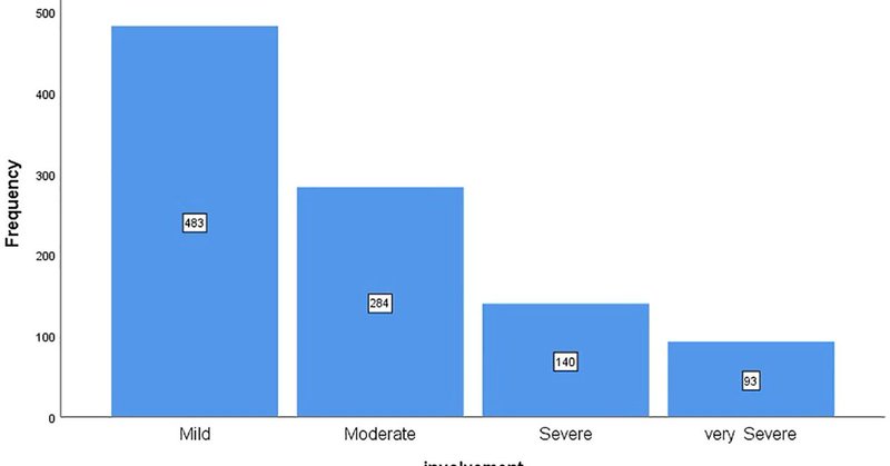
Most microadenomas were completely stable. Even in patients with 5+ years of imaging follow-up, there was no meaningful growth and no clinically relevant enlargement on serial MRI. This suggests that aggressive imaging protocols may not match real-world tumor biology.
0
0
0
Contrast-enhanced T1 had the highest detection rate (88.6%), but non-contrast sequences still detected a significant proportion (70.9% on T2W, 65.5% on T1W). Key question: if tumors aren’t growing, why do we perform routine contrast every year?
0
0
0
Are we overusing gadolinium in pituitary microadenoma follow-up? Our new study analyzing 300 MRIs shows that most microadenomas stay radiologically silent for years—and many are already detectable without contrast. https://t.co/H67N74Ecjl
6
1
0
Depression in RRMS has measurable MRI correlates—especially in limbic and insular regions—even in early, low-disability disease. Full article: https://t.co/r8VfwI0yIO
#MSResearch #Neuroimaging #Radiology #MentalHealthAwareness
0
0
0
BDI scores correlated with lesion load in mood networks: Left insular cortex: r = 0.20 Total insular cortex: r = 0.19 Even small disruptions in these hubs influence emotional symptoms. https://t.co/r8VfwI00Tg
0
0
0