
Stowers Institute Electron and Light Microscopy
@StowersMicro
Followers
104
Following
20
Media
7
Statuses
19
The support team for all manner of Microscopy at the Stowers Institute for Medical Research @ScienceStowers.
Kansas City, MO, USA
Joined September 2019
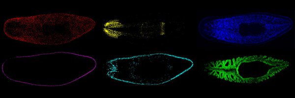
Great to see such beautiful work from the Gibson Lab including this gorgeous movie from our own @MelainiaM, @__ChrisWood__, and others from the microscopy core!!

In new work from our lab, @kzsabin explores the development and cell type composition of the Nematostella apical sensory organ (ASO), which is associated with the incredible apical tuft cilia (AT) shown in the attached movie. Check it out! https://t.co/nup2YpWxE0
0
2
7
Lots of really interesting neuro imaging coming our way working with the Özel lab! Glad to have you here Neset!

“The Özel Lab at the Stowers Institute will be an interdisciplinary team of computational and experimental biologists, working together with the singular goal of understanding brain development.” Learn more about one of our newest investigators, Neşet Özel https://t.co/7FSfJunvVk
0
3
9
Everyone around here is looking forward to imaging a lot of new things with @SivaSankariIMS, Neset Özel, and @MashruwalaAmeya!
The Stowers Institute is proud to announce the appointment of 3 new investigators. @SivaSankarilMS, PhD, Neset Özel, PhD, and @MashruwalaAmeya, PhD join the Institute to further advance our mission of scientific research benefitting humanity. Learn more:
0
2
9
We are really excited to get to work with @SivaSankariIMS and her lab to help answer fundamental questions about the relationship between bacteria and plants!
“The highly interdisciplinary, collaborative structure of the Stowers Institute is an ideal place to answer my complex #research questions." Learn more about one of our newest investigators, @SivaSankariIMS. https://t.co/hVAd3qBdmW
1
1
8
#MaintenanceMonday today is cleaning and getting some cups ready for a round of high-pressure freezing followed by freeze substitution with @MelainiaM and @S_Nowotarski
0
4
15
This week's stunning #MicroscopyMonday is sweet taste receptor neurons in the fruit fly proboscis. Image author: @Cheliuzz (Si Lab) #innovation #discovery
0
4
22
Listen as @Vidyanandsasi explains how when Stowers #postdocs have a very creative experiment in mind, our Technology Centers help make it happen. Learn more about our #Postdoc Training Program: https://t.co/ttGH3viesl
#TechEDGE
0
3
10
What is a Technology Center at Stowers? Listen as #postdoc @fg_mann, @Planaria1 Lab, explains more about these shared and collaborative facilities staffed with experts. https://t.co/ttGH3viMhT
#biology #innovation #TechEDGE
0
4
17
#MaintenanceMonday around here today. We are checking our diamond microtome knives for scratches by cutting empty resin. There's nothing worse than hitting the right spot in a sample and the wrong spot on your knife! Photo and checking by @MelainiaM
0
3
17
#WhyStowers? Investigator positions are fully funded and full of possibility. Hear our scientists explain why doing science at Stowers is different. Apply now to join our team as we pursue and discover the secrets of life. https://t.co/dyZCSG2LMB
#wearehiring #hiring #biologyjobs
1
38
112
The new Zeiss Elyra 7 Lattice SIM is installed and ready for imaging! Come by 454 to chat about what types of questions we can answer for you with this new technology!
1
5
37
The Opera Phenix is giving the green light to experiments in its new home on the 4th floor!
0
1
22
This is our favorite experiment from this paper. Transient optogenetic control of one protein causes long term aggregation of another! Wow! #optogenetics #Microscopy
The latest paper from @HalfmannLab is now fully formatted at @eLife, https://t.co/Ej8wvGCmfU. In the video, @arg_arginine uses a laser pulse to create “seeds” of CARD9 (top) that trigger activation of BCL10, how our #ImmuneSystem responds to pathogens. @unruhjay @jeff_j_lange
0
2
5
This video comes from @ahmetckarabulut in @Gibson_Lab in collaboration with @MelaniaM and shows the organelle responsible for sea anemone stinging using #volumeEM (SBF-SEM), rendered with @Blender. Read more about its mechanism for stinging here! https://t.co/rXLEXqm5gM
1
8
28
Video of a mouse brain shows how our #microscopy and #histology teams employ chemical clearing techniques & immerse samples in special solutions eliminating distortions. This gives a clear picture of the biological signals from complex tissues. 📹 author: Jeff Lange
0
1
15
It was great to have the opportunity to show the mayor what we can do in our wonderful facility with @phd_sarah and @MelainiaM!

My thanks to all at the @ScienceStowers for bringing phenomenal scientists and academics to our Greater Kansas City community and making our world a smarter, safer, and healthier place.
0
3
11
Our own Sarah Smith has spent some of her extra time putting together a tutorial on scientific figure preparation, check it out here: https://t.co/FExxZY6xYQ
youtube.com
This course is focused on scientific figures preparation using Adobe Illustrator and Fiji.
0
3
12
It’s alive! Keep an eye on your power generators: AT-AT, our new Nikon spinning disk, is available for usage. AT-AT is very similar to 3PO except that it has a second set of lasers for doing simultaneous imaging and bleaching. More info on Helix.
1
6
27