
Pere Roca-Cusachs
@PCusachs
Followers
1K
Following
201
Media
19
Statuses
215
Biophysicist at @IBECbarcelona, @unibarcelona.
Barcelona, Espanya
Joined October 2019
#PCBCommunity | An @IBECBarcelona-led study reveales how #mesenchymal_stem_cells respond to viscosity in their environment 📝@NatureComms The findings could revolutionise the #biomaterials design for #regenerativemedicine ▶️ https://t.co/YkFUTZlq4j
@ManuelMime @ribonucleico3
0
2
7
The system is adaptable to different cell types, different coatings, and single/multiple cell settings, so stay tuned for more on this in the future!
0
0
3
The result of hard, systematic, and precision work over the years by Laura Faure (experimental) and Manu Gómez (computational), in a joint effort between the labs of @XavierTrepat and our own. In collaboration with the labs of Marino Arroyo (UPC) and Elena Martínez (IBEC).
1
0
6
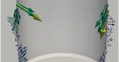
This allows us to average forces across multiple cells with the same shape, unveiling patterns in different directions. We show that as cell volume is reduced, epithelial cells transition from contractile to extensile forces!
1
0
3
There are available techniques to control cell shape in 3D, and to measure forces in 3D. But not to combine both! We achieve this by developing a 3D traction force microscopy method applied to cells micropatterned on polyacrylamide microwells.
1
0
5
Our work on a new technique we call 3D micropatterned traction force microscopy (3D-μTFM) is now out: https://t.co/n6nTjrTtyS
advanced.onlinelibrary.wiley.com
The function, mechanics, and 3D shape of cells are related to each other. Here, a technique is developed to control 3D cell shape, while measuring the mechanical forces that cells exert on their...
2
24
66

Thrilled to share my postdoctoral work exploring how brain mechanics influences metastasis, now out in @NatureCellBio. Check it out! #ChenLab @BUmechanobio @BioDesignCenter @wyssinstitute
https://t.co/DqmpleGpEq
nature.com
Nature Cell Biology - Uroz et al. report that the distinct mechanical properties of brain vasculature versus parenchyma drive cancer cell migration through a talin-dependent mechanism, enabling...
18
59
254

Through 3D micropatterned traction force microscopy, scientists can now measure the forces cells exert on their environment. @PCusachs and team used it to show how epithelial cells switch between contracting and extending as their volume changes. 👉 https://t.co/KsQuAo6FCF
0
2
12
.@i_GraneroMoya, @AndreuIon, @PCusachs & colleagues find that nucleocytoplasmic transport senses mechanical forces independently of cell density in cell monolayers. #OpenAccess #ReadAndPublish Highlight: https://t.co/aULD3nULD7 Article: https://t.co/HuSlXhjTNR
2
5
17

Looking for a summer read?🏖️🌞📄 You're in luck 😂. Our paper on optogenetic leader cells is out @NaturePhysics. We found a force-velocity relation for collective cell migration. Experiments by @LeoneRossetti, theory by @ricardalert. @IBECBarcelona
https://t.co/eAxUVIDs5P
5
51
250
The result of hard, systematic, and precision work over the years by Laura Faure (experimental) and Manu Gómez (computational), in a joint effort between the labs of @XavierTrepat and our own. In collaboration with the labs of Marino Arroyo (UPC) and Elena Martínez (IBEC).
2
0
1
This allows us to average forces across multiple cells with the same shape, unveiling patterns in different directions. We show that as cell volume is reduced, epithelial cells transition from contractile to extensile forces!
1
0
0
There are available techniques to control cell shape in 3D, and to measure forces in 3D. But not to combine both! We achieve this by developing a 3D traction force microscopy method applied to cells micropatterned on polyacrylamide microwells.
1
0
1
Here is our new pre-print on a new technique we call 3D micropatterned traction force microscopy (3D-μTFM): https://t.co/h7Sq9zNn0F

biorxiv.org
Cell shape and function are intimately linked, in a way that is mediated by the forces exerted between cells and their environment. The relationship between cell shape and forces has been extensively...
2
23
79
Painstaking, thorough, beautiful work by Srivatsava Viswanadha,recent Ph.D. graduate in my lab. Work co-directed between @ZanettaKechagia and myself, In collaboration with @KevinChalut and @XavierTrepat labs, with contributions by Manu Gómez, @celinelabo , and @valeria_ventur.
0
0
7
We show that forces increase during, and are required for, naïve pluripotency loss. This is mediated by GSK3 signalling. As a teaser, here is a naive pluripotency marker (Rex1-GFP, left), and traction forces (right) for a cell colony losing pluripotency (bottom) or not (top)
1
0
9
The very first step in the differentiation of mouse embryonic stem cells is the loss of naïve pluripotency. This step is accompanied by adhesion to the extracellular matrix, which typically involve cell-matrix force transmission. However, what is the role of these forces?
1
0
1
With a bit of delay in announcing it, here is our new preprint on the role of force in the loss of naïve pluripotency: https://t.co/OikS5bt6Fx
biorxiv.org
The first step in the differentiation of mouse embryonic stem cells (mESCs) is the loss of naïve pluripotency. This step involves a major reshaping of cells from a rounded to a spread, adhesive...
2
22
96